Evolution of the sphingolipid biosynthetic pathway
Sphingolipids are unique among eukaryotic cell lipids inasmuch as their biosynthesis is compartmentalized between the endoplasmic reticulum (ER) and the Golgi apparatus. This compartmentalization was first recognized about thirty years ago, and the lab is currently studying the ramifications of this feature for our understanding of how the pathway could have evolved. Studies over many decades have identified the enzymes in the pathway, their localization, topology, and an array of regulatory mechanisms. However, little is known about the evolutionary forces that underlie the generation of this complex pathway. We are currently studying the mechanisms by which the enzymes of the sphingolipid biosynthetic pathway are targeted to their different subcellular locations, how the pathway per se may have evolved, including its compartmentalization, and the relationship of the pathway to eukaryogenesis. Our studies have led to the suggestion that current models to describe evolution of the sphingolipid biosynthetic pathway may need substantial refinement.
It has been suggested that this approach is ‘obstructionist'; after all, we are here, so we must have gotten here somehow, and this must have involved an evolutionary process of some kind. While the latter is absolutely true, it does not address the details of how these evolutionary events could have occurred, and the details in the case of the SL biosynthetic pathway are perplexing to say the least.
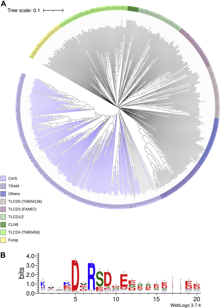
A. Phylogenetic tree showing the relationship between proteins with the TLC domain. The CerS clade is colored light blue. B. The DxRSDxE motif from the CerS branch of the tree. The DxRSDxE motif was shown to differentiate the CerS clade from the rest of the tree, as well as to be involved in CerS dimerization. It remains unclear both how this sequence and its function evolved, and additionally how redundant the sequence is.
Figure taken from: Kim J. L., et al. A novel C-terminal DxRSDxE motif in ceramide synthases involved in dimer formation. J. Biol. Chem. 298, 101517 (2022). https://doi.org/10.1016/j.jbc.2021.101517.