Gur Lab


Research
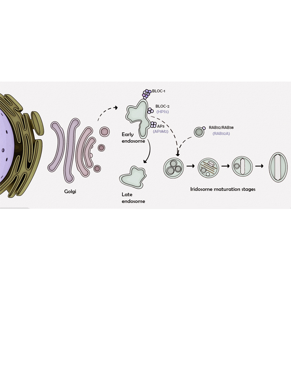
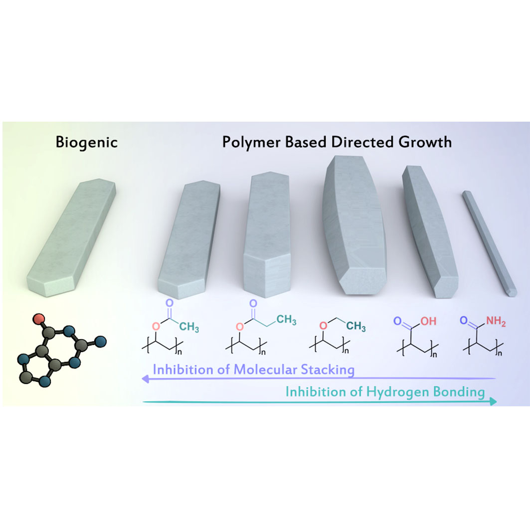
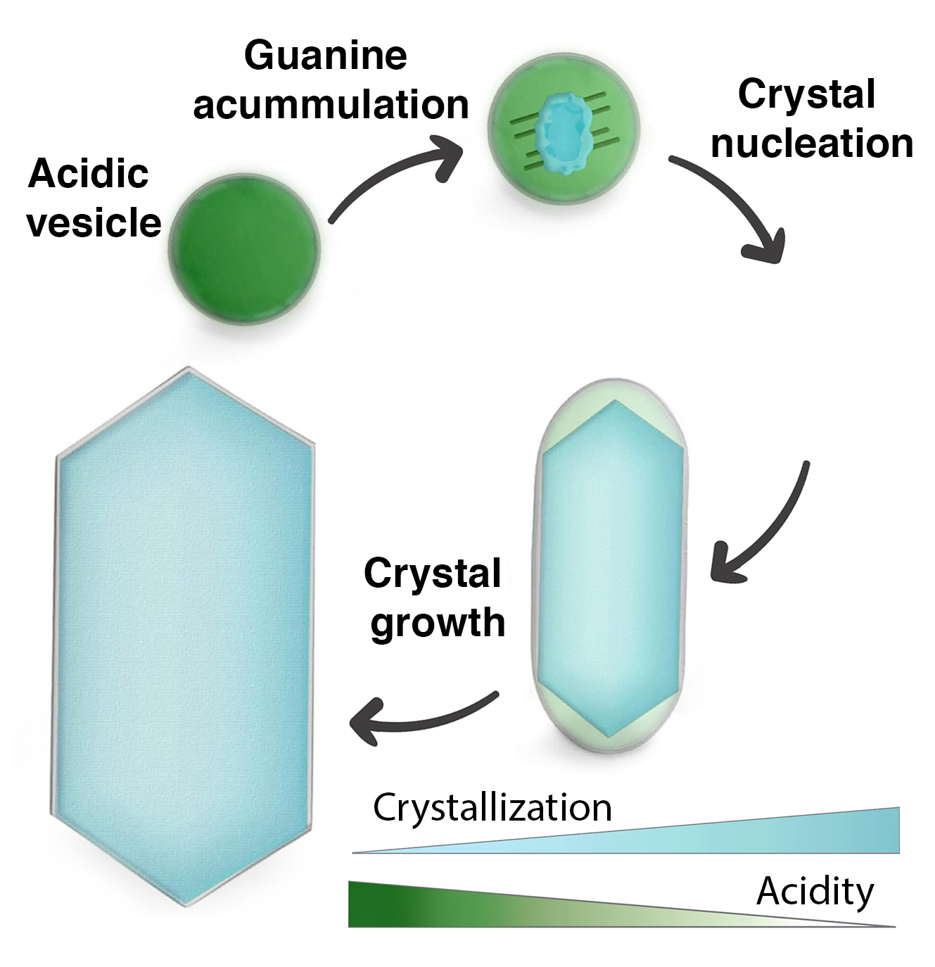
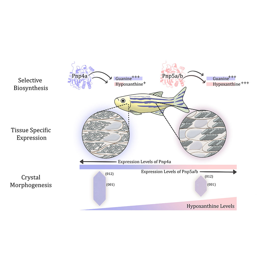
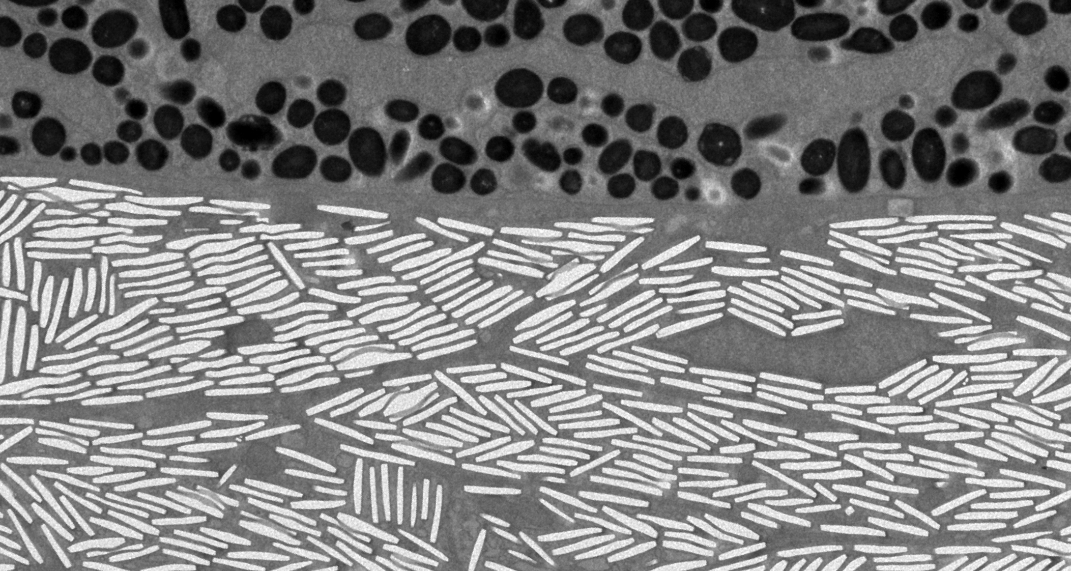
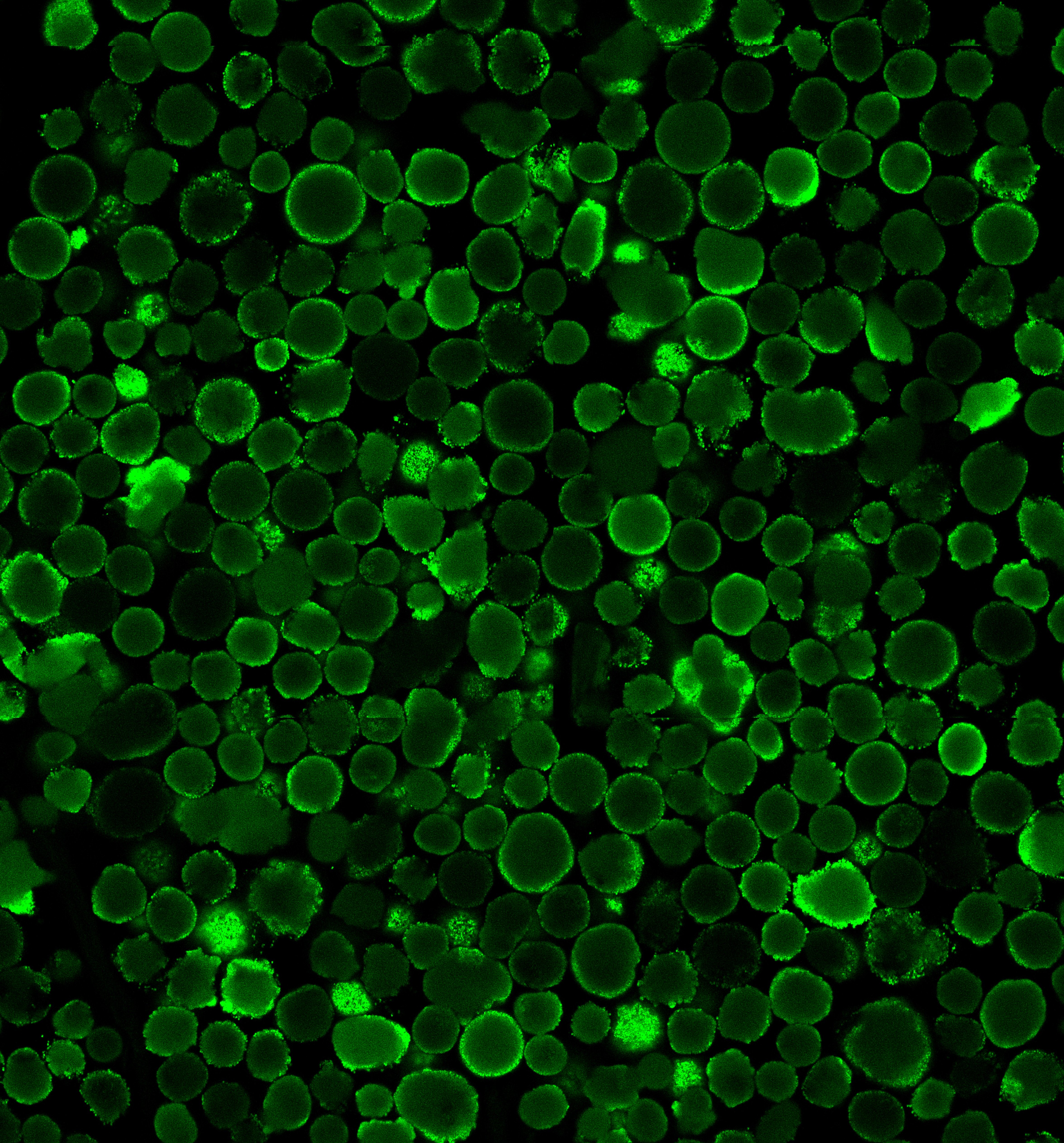
Chameleons, copepods, fish, and various other organisms employ organic crystals for an incredible array of optical purposes. These specialized crystals are produced by specific cells that exhibit unparalleled control over crystal shape, size, and assembly, surpassing even the most advanced techniques in materials science. Despite the discovery of these cells many years ago, very little is currently understood about their biology, specifically the cellular mechanisms responsible for organic crystal formation. To address this knowledge gap, we leverage a range of biological, physical, and chemical techniques to investigate the processes involved in the formation of both optically functional and pathological bio-organic crystals. Our research primarily focuses on zebrafish and medaka as model organisms.
News

Yael is the recipient of the The ISM Travel Award to MSC 2026!