
Method Specification
With the exception of the environmental SEM, liquid water cannot be observed in the SEM due to the high vacuum in the microscope’s column. Due to this limitation, most aqueous samples, biological or synthetic, are observed in the SEM only after the application of chemical fixation and dehydration procedures, which may cause severe alteration to the ultrastructure of the sample.
The frozen state of water is, however, compatible with vacuum in the SEM. In order to observe frozen samples the SEM should be equipped with a stable cold or cryo stage, which can keep the sample at a low temperature. The sample itself should be frozen without allowing water in the sample to crystalize. This can be achieved either by plunge freezing into liquid ethane when the sample is thin (up to few µm ) or by high pressure freezing when the sample is thicker (~200 µm). SEM allows only for the observation of the sample’s surface. In order to observe inner parts of the sample, the frozen sample can be fractured under vacuum in a freeze fracture device (see figure, upper panel). The freshly exposed surface can also be coated with a thin layer of metal or carbon in order to enhance contrast and reduce damage by the electron beam in the microscope.
Interpretation of cryo SEM images formed by secondary electrons is not always straightforward, unlike in TEM imaging, contrast is not formed by density differences (i.e. membranes are darker than the cytoplasm), but by the topography of the surface (see figure, lower panel). When a back-scattered electrons (BSE) detector is used, cryo SEM images can also provide information on the consistency of the sample as elements with high atomic number would be brighter than lighter elements. While the chemical identity of the observed structure cannot be discerned, it is sometimes sufficient to recognize regions containing heavier elements on the background of lighter ones. For instance calcium on the background of carbon and oxygen can provide information on the localization of mineralized regions in bones. In order to identify elements unequivocally, characteristic x rays emitted from the sample upon interaction with the electron beam can be measured. The energy of the emitted x ray photon can be related directly to the element that released this photon.
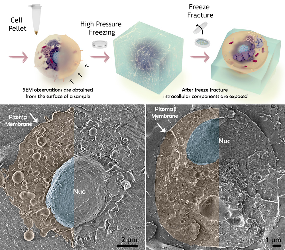
Cryo freeze fracture for SEM imaging. Upper panel shows schematically the conceptual idea behind the freeze fracture process. In order to observe inner parts of the sample, the frozen sample is fractured under vacuum in a freeze fracture device and the exposed surface is then explored under the electron beam. Lower panel shows two examples of freeze fractured cells. The cell cytoplasm is pseudo colored in yellow and the nucleus in blue.
Figure by Neta Varsano.
Staff Contacts
-
-

Dr. Neta Varsano
Staff Scientist