Method Specification
Cryogenic transmission electron microscopy is conducted on specimens that are cooled in the microscope. Cryogenic observation, such as at liquid nitrogen temperature, improves the stability of the material or allows for observing a material whose properties change at lower temperatures.
Modern automated cryo-TEMs are designed specifically to operate at liquid-nitrogen temperature. By combination with recent electron counting detectors that are sensitive to a single electron cryo-TEM became the method of choice for characterisation in structural biology, in many cases replacing x-ray diffraction for protein structure analysis.
Even though cryo-operation is less common in materials science, there are opportunities opening with new instrumentation to study materials that tend to damage fast under the electron beam or to take benefit of cryo-preservation to study the pristine state of materials instead of a possibly altered structure of dried products.

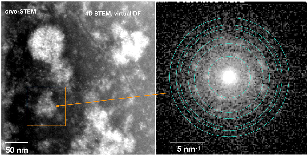
Figure: Scanning nanodiffraction of CaF2 growing from an intermittent aggregate formed in Ca2+/AEP solution after addition of NaF. The sample was prepared from the reactant solution by plunge-freezing. Pure phase CaF2 nanoparticles are forming and coalescing in the larger scale aggregates that consist of residual AEP competing with F over the Ca cations. At a later stage of the reaction CaF2 nanoparticles are released form the aggregates. (Mashiach et al. Nat Commun 12, 229 (2021). doi:10.1038/s41467-020-20512-6).
Further reading
- McComb, D.W., Lengyel, J. & Carter, C.B. Cryogenic transmission electron microscopy for materials research. MRS Bulletin 44, 924–928 (2019). doi: 10.1557/mrs.2019.283
Staff Contacts
-

Dr. Lothar Houben
Staff Scientist -

Dr. Olga Brontvein
Staff Scientist

