
Method Specification
Tomography aims at the reconstruction of the three-dimensional structure of a sample from two dimensional representations. The most common form of tomography in materials science is the reconstruction from a set of images of a single object taken under various viewing directions.
The basis of the reconstruction is the Radon transform that relates the three-dimensional space of image coordinates and the tilt angle to the three-dimensional cartesian space.
In practice tilt-series of images are taken and reconstructed with a backprojection algorithms. Complications arise from the so-called missing wedge of missing tilt angles through geometrical tilt restrictions in the TEM, insufficient alignment, sample transformations during tilt and failures in the projection requirement which states that the image intensity has to be a monotoneous function of mass density.
In materials science the most abundant form of tomography is STEM tomography, since it fulfils the projection requirement closer than TEM imaging that suffers from Bragg artefacts.
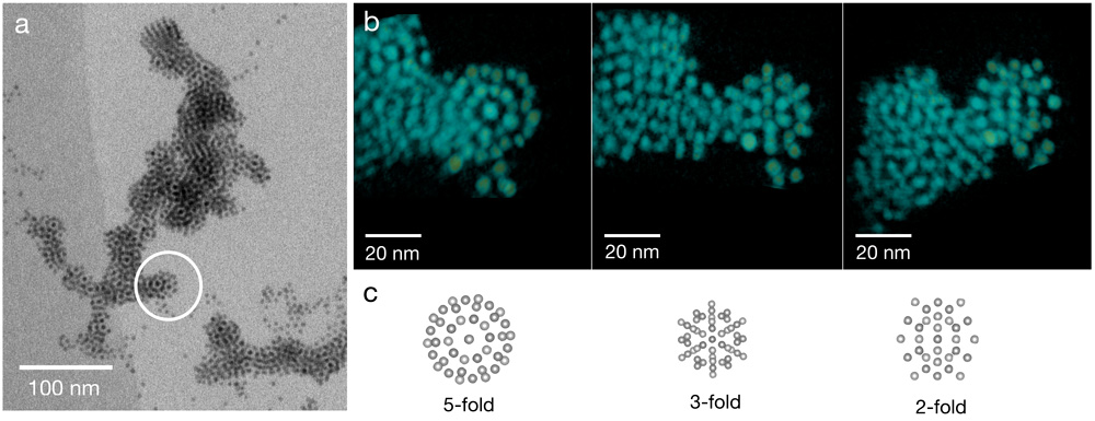
Figure: Icosahedral Mackay clusters in aggregates of Au NP. (a) Cryo-STEM bright-field image taken from a tilt series of images of the Au NP:STP aggregate. The circular marker exposes a cluster that is a building block of the aggregate. (b) Voxel projections of the tomographic reconstruction of the cluster marked in (a). Three projections corresponding to the 5-fold, 3-fold and 2-fold symmetry axes of the icosahedral symmetry are shown. (c) Projections of a matching two-shell Mackay cluster with a central NP, an inner icosahedral shell with 12 NP, and an outer deltahedral shell with 42 NP. (with: Tong Bian, Rafal Klajn, Dept. of Organic Chemistry, WIS).
Further reading
- Midgley PA, Weyland M. Ultramicroscopy. 2003 Sep;96(3-4):413-31. doi: 10.1016/S0304-3991(03)00105-0.
- Midgley PA, Dunin-Borkowski RE. Nat Mater. 2009 Apr;8(4):271-80. doi: 10.1038/nmat2406.
Staff Contacts
-

Dr. Lothar Houben
Staff Scientist -

Dr. Olga Brontvein
Staff Scientist