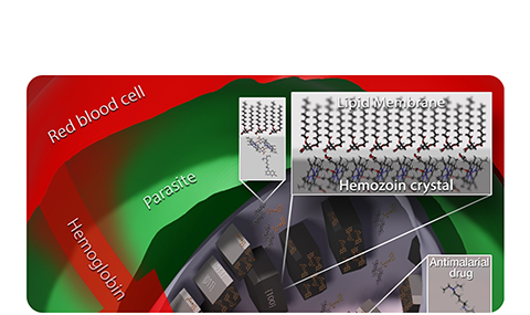
As already mentioned, crystallization of the malaria pigment (i.e., hemozoin) by the malaria parasite P. falciparum in infected human red blood cells is a crucial step for its survival. But this process is also the parasite’s Achilles’ heel since hemozoin is an antimalarial drug target. Over the past two decades, we have carried out a study on the nucleation, growth and detailed structure of hemozoin, via various experimental methods. These methods included, cryo soft X-ray tomography, X-ay fluorescence, cryo-electron diffraction tomography and X-ray powder diffraction. No less important, we also addressed the important issue of the mode of action of various antimalarials such as the quinolne and artemisinin drugs by a computational approach, and the quinoline drugs by cryo soft X-ray tomography and X-ay fluorescence. These drugs act by smothering the faces of hemozoin, not allowing the crystals to grow; the upshot of which is the perforation of the membrane by drug-hematin complexes, leading to parasitic death.
This work has been carried out with various students and collaborators, in particular Sergey Kapishnikov, Michael Elbaum and Jens Als-Nielsen. It has been recently reviewed in the following article by:
S. Kapishnikov, E. Hempelmann, M. Elbaum, J. Als-Nielsen, L. Leiserowitz, Malaria Pigment Crystals, the Achilles Heel of the Malaria Parasite, ChemMedChem 2021, 16, 1515–1532.


