Major league magnets
Nuclear magnetic resonance in the service of science
Features
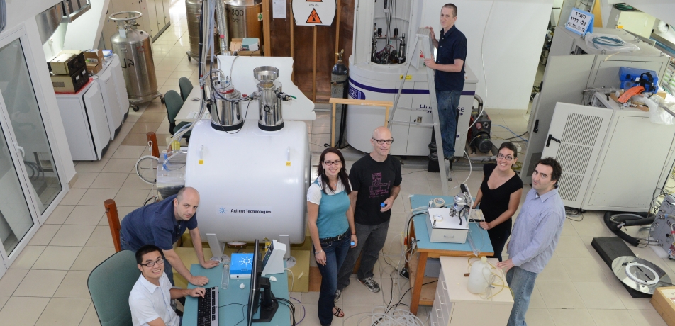
When the Weizmann Institute’s first Nuclear Magnetic Resonance (NMR) spectrometer was built at the Institute in the 1950s, it was an ingenious machine, cobbled together out of electronic equipment left behind by the British army, for the purpose of peering into a multitude of materials.
Later, NMR machinery was bought from manufacturers, but Weizmann Institute scientists have continued improving the equipment and refining the techniques, several of them becoming leaders in the field.
The Institute’s NMR activities span a wide range of scientific disciplines, ranging from physics and chemistry to cancer research and neurobiology. Much of this work has been funded through the Ilse Katz Institute for Material Sciences and Magnetic Resonance Research and the Helen and Martin Kimmel Institute for Magnetic Resonance Research. To stay on the cutting edge, the Institute is now planning on installing new scanners, including one that boasts 30 times the field of that original NMR - 21 Tesla - as well as an additional 7 Tesla NMR imaging (MRI) magnet that will be used on humans. (The numbers are a measure of magnetic strength; magnets used in clinical MRIs are 1.5-3 Tesla, for instance).
The first of these instruments will help Institute researchers, including a new generation of Weizmann Institute scientists, to develop innovative technologies for human health, advanced materials and biomolecular research. The second, a 7 Tesla instrument—for which the Israeli government recently pledged $8 million out of the total $12 million price tag - will be devoted to cancer and brain research. The Institute plans on acquiring additional magnets in the future.
Prof. Lucio Frydman of the Department of Chemical Physics, who is leading the Institute’s efforts in this realm, describes the basis of NMR and its familiar form, MRI—as centered on “quantum compass needles.” At the heart of any NMR scanner is a strong magnet that uses the magnetic properties of atomic nuclei to “polarize” them—that is, to align them parallel to the external magnet. When these aligned nuclei - for instance, the protons in water molecules that make up our bodies—are pulled away from their normal balance, they return to equilibrium, emitting low-energy radio waves in the process that can be used to characterize their position or their chemical environments.
But even with the strongest magnets, NMR technology has been hobbled by twin limitations: time and sensitivity. As anyone who has held perfectly still for an MRI knows, the process can be time-consuming. And the sensitivity of human scanners is usually only sufficient to image the water that makes up most of our body. Other types of NMR scans, for example those of complex molecules, would take hours or days, while many fragile or dynamic objects cannot be scanned at all by standard methods.
Prof. Frydman has developed “ultrafast” NMR and MRI methods, which can obtain multidimensional data and images in a fraction of a second. These methods have had most of their applications in the realms of chemistry, biology, and medicine. In studies conducted this past year together with Prof. Michal Neeman of the Department of Biological Regulation, ultrafast MRI equipment was used to image one of the most complex fluid flows in nature - the interface between fetal and maternal blood in the placenta. The ability to sort and image these entangled flows could be used in the future to identify early signs of fetal distress. Some of Prof. Frydman’s methods are already used in humans, including ongoing collaborative efforts with the group of Prof. Hadassah Degani of the Department of Biological Regulation to detect breast cancer without the use of contrast material.
Another breakthrough that Prof. Frydman is developing together with his Weizmann Institute colleagues Profs. Shimon Vega and Daniella Goldfarb of the same department concerns so-called hyperpolarization methods. This team aims to depart from the nuclear polarizations that conventional magnets can deliver - which are on the order of one nucleus out of every 100,000 becoming aligned—and orient nearly one out of every five “compass needles.”
This advance could open the door to imaging many kinds of molecules whose signals would normally be too faint to see in ordinary MRI. These include, for example, such metabolites as the lactate produced in leg muscles during running, or others whose levels are altered in the brain following a stroke. In recent studies, Prof. Frydman demonstrated the imaging of each of these in rodents. If they are eventually adapted for humans, the former could tell athletes whether their muscles are more evolved for “sprinting” or “distance running”; the latter could provide a non-invasive way to accurately assess which areas of the brain are affected by a stroke.
“This is what is so fascinating about research in magnetic resonance,” says Prof. Frydman. “It is a blend of theories and experiments deeply rooted in the elusive concepts of quantum physics, which demand overcoming significant engineering and signal processing challenges to yield their full potential. Yet as a result of these multidisciplinary efforts emerge real-life tools that, if blended with sufficient ingenuity, can provide us with unprecedented descriptions of dynamic molecular and biological processes, as well as with diagnostic evidences that can literally change the fate of individuals.”
Several new scientists have joined the ranks of the Weizmann Institute in this field in recent years (see sidebars). In September, Dr. Michal Leskes will join the Department of Materials and Interfaces, where she will focus on advancing technology for rechargeable batteries so they have faster charging capabilities and improved power storage. This work requires the ability to investigate lithium-ion and lithium-air batteries by monitoring the dynamics of electrochemical products formed at their electrodes during charging and discharging. This research will advance the overall effort in creating renewable energy options that are practical, dependable, and affordable.
Also, Dr. Rina Rosenzweig will join the Department of Structural Biology at the Weizmann Institute starting in 2016. She uses NMR to understand the molecular mechanisms involved in diseases associated with protein misfolding and the accumulation of toxic protein aggregates. These include Alzheimer’s and Parkinson’s diseases, type II diabetes, and the spongiform encephalopathies, such as Creutzfeldt- Jakob disease. Therefore, understanding the molecular mechanism involved in triggering protein aggregation in vivo is essential to prevent, slow down, or even ultimately, reverse the progression of these diseases.
Dr. Assaf Tal: Beyond the standard fMRI
Dr. Assaf Tal works between two worlds: One is that of subatomic particles exposed to strong magnetic fields and the other, no less mysterious, is the human brain. His office is in the Department of Chemical Physics; his experiments are conducted in the fMRI lab in the Arison Laboratory for Human Brain Imaging.
Dr. Tal, who grew up in Rehovot, completed an MSc in the Weizmann Institute group of Prof. Gershon Kurizki in Chemical Physics and did his PhD research in the lab of Prof. Frydman. After a short stint in industry, Dr. Tal returned to postdoctoral research, first in the lab of the Institute’s Prof. Hadassa Degani and then at New York University Medical Center, at a radiology lab that specializes in imaging the brain.
His decision to return to Israel, he says, was dependent on a magnet, specifically the super-strong magnet found in MRI machinery: “Few research institutions could offer me both access to a large magnet and openness to collaboration with neurobiologists. Here, at the Weizmann Institute, I am building that connection.”
Dr. Tal is attempting to image such large biomolecules as neurotransmitters— substances used by neurons to communicate. His challenge is to separate out the signal of these from that of the ubiquitous water. “It’s like training the ear to hear the notes played by just one instrument in an entire symphony,” says Dr. Tal.
Imaging neurotransmitters might enable researchers to observe such elusive processes as the consolidation of memories. “We want to move beyond the standard fMRI and ask totally new questions about the workings of the brain,” says Tal.
Dr. Amnon Bar-Shir: In living color
Dr. Amnon Bar-Shir has a dream. In it, today’s black-and-white MRI technology will be brought to us in living color. Those colorful images will greatly increase our ability to observe and trace, for example, the success of implanted cells, the progress of anti-cancer treatments, or the release of neurotransmitters in the brain.
The inspiration for some of his work comes directly from the fluorescent protein technique that has revolutionized microscopy and for which the Nobel Prize in Chemistry was awarded in 2008. “Why not do the same thing for MRI?” says Dr. Bar-Shir, who joined the Weizmann Institute in 2014 after a postdoctoral fellowship at Johns Hopkins University School of Medicine. “Then we could observe many processes deep inside tissues of live subjects, non-invasively and over time.” Dr. Bar-Shir has been working on developing proteins that may yield different colors in the MRI scanner. He helped create a viral protein that, when paired with a synthetic sensor, acted as a high-contrast, colorful reporter in a mouse brain.
“I hope to create a whole palette of colors,” he says. “The idea is that we can image several different molecules at once. If we insert different color-coded genes into immune cells that have been modified outside the body to fight cancer, for example, we might be then able to see where these cells actually go in the body and how they attack the tumor cells. In the future, this might be used as a diagnostic test to fine-tune treatment.”
Major government investment in brain imaging
In December the Israeli government’s Forum for National Infrastructure Forum for Research and Development (TELEM), which is tasked with budgeting for major national science and technology initiatives - announced its decision to provide major funding to establish the Israel National Center for Brain Imaging and Stimulation led by the Weizmann Institute of Science and in collaboration with the Technion - Israel Institute of Technology, the Hebrew University of Jerusalem, and the Tel Aviv Sourasky Medical Center. The Center will be located on the Weizmann Institute campus and will revolve around the facility.
“This decision is a huge vote of confidence by the Israeli government for the Weizmann Institute,” says Prof. Daniel Zajfman, President of the Weizmann Institute. The joint proposal submitted to TELEM was reviewed by experts from abroad who said that the “excellent” project “makes considerable sense given the Israeli research landscape in human cognitive neuroscience… It will bring Israel to an even higher level of brain imaging capacity, available to only the best international neuroscience centers.”
Telem will allocate millions of dollars for the establishment of the Israel National Center for Brain Imaging and Stimulation, to be divided between the Weizmann Institute (which will receive the lion’s share), Technion, and Sourasky Medical Center. The funds provided by the Israel government to the Weizmann Institute will cover the cost of purchasing the system. Each of the collaborating entities in the Israeli National Center for Brain Imaging and Stimulation will provide an additional aspect to brain imaging.
Prof. Lucio Frydman is funded by Adelis Foundation, The Clore Institute for High-Field Magnetic Resonance Imaging and Spectroscopy, European Research Council, Paul and Tina Gardner, Austin TX, Leona M. and Harry B. Helmsley Charitable Trust, The Ilse Katz Institute for Material Sciences and Magnetic Resonance Research, Helen and Martin Kimmel Award for Innovative Investigation, The Helen and Martin Kimmel Institute for Magnetic Resonance Research which he heads, Gary and Katy Leff, Calabasas, CA, The Mary Ralph Designated Philanthropic Fund of the Jewish Community Endowment Fund, Takiff Family Foundation. Prof. Frydman is the incumbent of the Bertha and Isadore Gudelsky Professorial Chair.
http://www.weizmann.ac.il/chemphys/Frydman_group
Dr. Assaf Tal is funded by Carolito Stiftung, Monroy- Marks Career Development and Staff Scientist Start-Up Fund, Leona M. and Harry B. Helmsley Charitable Trust. Dr. Tal is the incumbent of the Monroy-Marks Career Development Chair.
http://www.weizmann.ac.il/chemphys/assaf_tal/
Dr. Amnon Bar-Shir is funded by The Benoziyo Fund for the Advancement of Science, The Willner Family Leadership Institute for the Weizmann Institute of Science, The Gurwin Family Fund for Scientific Research.
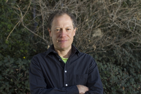
Prof. Lucio Frydman
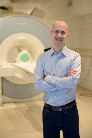
Dr. Assaf Tal
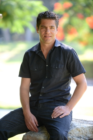
Dr. Amnon Bar-Shir


