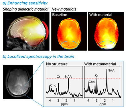
There is a continuous effort to improve the signal in MRI, especially vital while developing new biomarkers and achieving higher resolution. In addition, as the magnetic field increases the RF wavelength inside the body decreases. Once the wavelength is of the order of the object/region of interest, local interferences appear, also called wavelength effects1,2. These interferences generate local shading which are significant at 7 T in almost any part of the body, including brain imaging. Dielectric materials are already in common use as pads attached to the region of interest, for example for brain imaging at 7T in the Human Connectome Project3. Studying specially designed artificial materials or also called metamaterials (combining conducting strips and high permittivity dielectric material), we can reach new capabilities of enhancing local sensitivity of MRI signal and resulting imaging resolution.
Inspired by the development of artificial materials in the RF fields, such as optics, microwave and acoustics, we adapt the metamaterial concepts to MRI and explore new MRI-viable metamaterials, taking into account required considerations. These include constraints due to the RF frequencies involved, patient environment (bearing in mind patient comfort) as well as spin excitation and signal reception from deep regions in the body, which is essential for in-vivo imaging covering all organs. Moreover, one of the crucial prerequisites is delivering the required tailored RF field within safe levels of the power deposition in the body.

Dielectric and metamaterial’s simulation of EM fields distributions and in-vivo localized 1H spectroscopy. a, RF simulations example using dielectric and metamaterials for local signal enhancement. b, Localized 1H spectroscopy performed without (left) and with (right) metamaterial pad.
1. Collins, C. M., Liu, W., Schreiber, W., Yang, Q. X., & Smith, M. B. (2005). Central brightening due to constructive interference with, without, and despite dielectric resonance. Journal of Magnetic Resonance Imaging, 21(2), 192-196.
2. Vaughan, J. T., Garwood, M., Collins, C. M., Liu, W., DelaBarre, L., Adriany, G., ... & Ugurbil, K. (2001). 7T vs. 4T: RF power, homogeneity, and signal‐to‐noise comparison in head images. Magnetic resonance in medicine, 46(1), 24-30.
3. Vu, A. T., Auerbach, E., Lenglet, C., Moeller, S., Sotiropoulos, S. N., Jbabdi, S., ... & Ugurbil, K. (2015). High resolution whole brain diffusion imaging at 7 T for the Human Connectome Project. Neuroimage, 122, 318-331.


