Publications
Steffen Jung's complete bibliography
2026
-
(2026) Encyclopedia of Immunobiology. 2nd ed. p. V1:45-V1:51 Abstract
Macrophages are critical players in physiology and pathophysiology, and an in-depth understanding of their diverse origins and specific functions holds promise for therapeutic intervention. Mammalian macrophages can arise from consecutive waves of hematopoietic development. Two of these pathways that commence in extra-embryonic tissue remain confined to the fetus and cease before birth, although some of their macrophage products persist throughout life. The third wave of macrophages derives from hematopoietic stem cells (HSC) that arise in the embryo proper and sustain lifelong myeloid hematopoiesis. These latter cells rely on blood monocytes for dissemination and progressively replace existing macrophage compartments to a varying extent. In this chapter, we discuss the current understanding of macrophage ontogeny and its physiological impact and promise for therapeutic strategies.
(2026) Nature Immunology. 27, 1, p. 2-3 AbstractThe array of tissue macrophages characterized is ever-expanding, underscoring the importance of these cells in physiology and pathology. A population of specialized gland-associated macrophages termed adenophages has now been discovered that support efficient saliva secretion along with possible other immunological functions.
2025
-
(2025) European Journal of Immunology. 55, 11, e70077. Abstract[All authors]
The timing of endotoxin administration in mice matters and is associated with diurnal variation in survival; however, underlying mechanisms remain poorly understood. Here, we report that afternoon LPS challenges in mice induce a robust inflammatory response involving increased neutrophil activation and release of cytotoxic mediators, causing higher mortality compared with challenges at midnight. Mechanistically, the cyclic patterns of corticosterone and melatonin hormones differentially modulate neutrophil responses. The afternoon corticosterone peak was associated with heightened incidence and severity of LPS-induced hyperinflammation. Conversely, higher melatonin levels at midnight conferred protection to challenged mice by restraining the magnitude of inflammation. High cortisol and low melatonin profiles detected in septic patients mirror those observed in mice and suggest a novel prognostic marker for sepsis. Our study unveils a regulatory network that links light/dark signals and circadian-regulated hormones to the intensity of the host's inflammatory response to infection.
(2025) Reference Module in Life Sciences. Second ed. AbstractMacrophages are critical players in physiology and pathophysiology, and an in-depth understanding of their diverse origins and specific functions holds promise for therapeutic intervention. Mammalian macrophages can arise from consecutive waves of hematopoietic development. Two of these pathways that commence in extra-embryonic tissue remain confined to the fetus and cease before birth, although some of their macrophage products persist throughout life. The third wave of macrophages derives from hematopoietic stem cells (HSC) that arise in the embryo proper and sustain lifelong myeloid hematopoiesis. These latter cells rely on blood monocytes for dissemination and progressively replace existing macrophage compartments to a varying extent. In this chapter, we discuss the current understanding of macrophage ontogeny and its physiological impact and promise for therapeutic strategies.
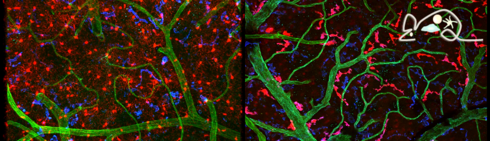
(2025) Proceedings of the National Academy of Sciences - PNAS. 122, 21, e241431612. AbstractConcussions can cause debilitating symptoms despite no evidence of structural changes on diagnostic imaging. The cellular events occurring in the brain parenchyma following concussion, especially repetitive concussion, are not well elucidated. We developed a concussion model to induce a confined area of injury without causing frank hemorrhage. Using intravital microscopy, we observe activation of the vasculature that supported neutrophil rolling and platelet adhesion but no overt cellular recruitment from blood into brain parenchyma. Activated resident, not monocyte-derived, macrophages relocated to the injury site via Cx3cr1 and phagocytosed dysfunctional/ detached astrocytes via scavenger receptors and TLR4, particularly after repetitive concussion. Additionally, microglia sealed areas of bloodbrain barrier (BBB) disruption via purinergic pathways. Using a splitCre approach to dissect microglia and perivascular macrophages, we show that microglial invasion into the injury site is key to reducing BBB disruption. Our data suggest that microglia repair the BBB following concussion, but in doing so significantly alter the cellular ultrastructure of the brain milieu.
(2025) Cell Reports. 44, 5, 115609. Abstract[All authors]Summary Microglia are parenchymal brain macrophages that are established during embryogenesis and form a self-containing cellular compartment that resists seeding with cells derived from adult definitive hematopoiesis. We report that monocyte-derived macrophages (MoMΦs) accumulate in the brain of aging mice with distinct topologies, including the nigrostriatum and medulla but not the frontal cortex. Parenchymal MoMΦs adopt bona fide microglia morphology and expression profiles. Due to their hematopoietic stem cell (HSC) derivation, monocyte-derived microglia (MoMg) are unlike yolk-sac-derived cells, targets of clonal hematopoiesis (CH). Indeed, using a chimeric transfer model, we show that the hematopoietic expression of DNMT3AR882H, a prominent human CH variant, renders MoMg pathogenic and promotes motor deficits resembling atypical Parkinsonian disorders. Collectively, we establish that MoMg progressively seed the brain of healthy aging mice, accumulate in selected areas, and, when carrying a somatic mutation associated with CH, can cause brain pathology.
2024
-
(2024) Science immunology. 9, 99, eadp0344. Abstract[All authors]
Langerhans cells (LCs) are distinct among phagocytes, functioning both as embryo-derived, tissue-resident macrophages in skin innervation and repair and as migrating professional antigen-presenting cells, a function classically assigned to dendritic cells (DCs). Here, we demonstrate that both intrinsic and extrinsic factors imprint this dual identity. Using ablation of embryo-derived LCs in the murine adult skin and tracking differentiation of incoming monocyte-derived replacements, we found intrinsic intraepidermal heterogeneity. We observed that ontogenically distinct monocytes give rise to LCs. Within the epidermis, Jagged-dependent activation of Notch signaling, likely within the hair follicle niche, provided an initial site of LC commitment before metabolic adaptation and survival of monocyte-derived LCs. In the human skin, embryo-derived LCs in newborns retained transcriptional evidence of their macrophage origin, but this was superseded by DC-like immune modules after postnatal expansion. Thus, adaptation to adult skin niches replicates conditioning of LC at birth, permitting repair of the embryo-derived LC network.
(2024) Journal of Experimental Medicine. 221, 5, e20231686. Abstract[All authors]The mycobiota are a critical part of the gut microbiome, but hostfungal interactions and specific functional contributions of commensal fungi to host fitness remain incompletely understood. Here, we report the identification of a new fungal commensal, Kazachstania heterogenica var. weizmannii, isolated from murine intestines. K. weizmannii exposure prevented Candida albicans colonization and significantly reduced the commensal C. albicans burden in colonized animals. Following immunosuppression of C. albicans colonized mice, competitive fungal commensalism thereby mitigated fatal candidiasis. Metagenome analysis revealed K. heterogenica or K. weizmannii presence among human commensals. Our results reveal competitive fungal commensalism within the intestinal microbiota, independent of bacteria and immune responses, that could bear potential therapeutic value for the management of C. albicansmediated diseases.
(2024) Immunity. 57, 6, p. 1225-1242.e6 Abstract[All authors]Summary Classical monocytes (CMs) are ephemeral myeloid immune cells that circulate in the blood. Emerging evidence suggests that CMs can have distinct ontogeny and originate from either granulocyte-monocyte- or monocyte-dendritic-cell progenitors (GMPs or MDPs). Here, we report surface markers that allowed segregation of murine GMP- and MDP-derived CMs, i.e., GMP-Mo and MDP-Mo, as well as their functional characterization, including fate definition following adoptive cell transfer. GMP-Mo and MDP-Mo yielded an equal increase in homeostatic CM progeny, such as blood-resident non-classical monocytes and gut macrophages; however, these cells differentially seeded various other selected tissues, including the dura mater and lung. Specifically, GMP-Mo and MDP-Mo differentiated into distinct interstitial lung macrophages, linking CM dichotomy to previously reported pulmonary macrophage heterogeneity. Collectively, we provide evidence for the existence of two functionally distinct CM subsets in the mouse that differentially contribute to peripheral tissue macrophage populations in homeostasis and following challenge.
(2024) Microbiology Resource Announcements. 13, 4, e00115-24. AbstractKazachstania heterogenica is a member of the K. telluris complex, where all members to date are reported to be pathogenic fungi. We have isolated a strain, K. heterogenica var. weizmannii, from the gut of mice that seems to be a commensal strain and sequenced its genome.
(2024) Microglia. Verkhratsky A. & Tremblay M.(eds.). Vol. 37. p. 53-63 AbstractSince the classical studies of Pío del Río-Hortega, microglia research has come a long way. In particular, recent advances in bulk and single-cell (sc) transcriptomics have yielded many fascinating new insights into these intriguing immune cells at the interface with the central nervous system (CNS), both in small animal models and human samples. In parallel, tools developed by advanced mouse genetics have revealed the unique ontogeny of microglia and their striking dynamic interactions with other cells in the brain parenchyma. In this chapter, we will discuss various applications of the Cre/loxP-based approach that have enabled the study of microglia in their physiological context of the mouse brain. We will highlight selected key findings that have shaped our current understanding of these cells and discuss the technical intricacies of the Cre/loxP approach and some remaining challenges.
(2024) Tissue-Resident Macrophages. Mass E.(eds.). p. 253-267 AbstractGlobal gene expression profiling has provided valuable insights into the specific contributions of different cell types to various physiological processes. Notably though, both bulk and single-cell transcriptomics require the prior retrieval of the cells from their tissue context to be analyzed. Isolation protocols for tissue macrophages are, however, notoriously inefficient and, moreover, prone to introduce considerable bias and artifacts. Here, we will discuss a valuable alternative, originally introduced by Amieux and colleagues. This so-called RiboTag approach allows, in combination with respective macrophage-specific Cre transgenic lines, to retrieve macrophage translatomes from crude tissue extracts. We will review our experience with this ingenious method, focusing on the study of brain macrophages, including microglia and border-associated cells. We will elaborate on the advantages of the RiboTag approach that render it a valuable complement to standard cell sorting-based profiling strategies, especially for the investigation of tissue macrophages.
(2024) Tissue-Resident Macrophages. Mass E.(eds.). p. 481-503 AbstractMacrophages represent a broad spectrum of distinct, but closely related tissue-resident immune cells. This presents a major challenge for the study of functional aspects of these cells using classical Cre recombinase-mediated conditional mutagenesis in mice, since single promoter-driven Cre transgenic models often display limited specificity toward their intended target. The advent of CRISPR/Cas9 technology has now provided a time- and cost-effective method to explore the full potential of binary transgenic, intersectional genetics. Specifically, the use of two promoters driving inactive Cre fragments that, when co-expressed, dimerize and only then gain recombinase activity allows the characterization and manipulation of genetically defined tissue macrophage subpopulations. Here, we will elaborate on the use of this protocol to capitalize on these recent technological advances in mouse genetics and discuss their strengths and pitfalls to improve the study of tissue macrophage subpopulations in physiology and pathophysiology.
(2024) GLIA. 72, 1, p. 5-18 AbstractMacrophages have emerged as critical cellular components of the central nervous system (CNS), promoting development, maintenance, and immune defense of the CNS. Here we will review recent advances in our understanding of brain macrophage heterogeneity, including microglia and border-associated macrophages, focusing on the mouse. Emphasis will be given to the discussion of strengths and limitations of the experimental approaches that have led to the recent insights and hold promise to further deepen our mechanistic understanding of brain macrophages that might eventually allow to harness their activities for the management of CNS pathologies.
2023
-
(2023) Nature Neuroscience. 26, 3, p. 406-415 Abstract[All authors]
Alzheimers disease (AD) is characterized by synaptic loss, which can result from dysfunctional microglial phagocytosis and complement activation. However, what signals drive aberrant microglia-mediated engulfment of synapses in AD is unclear. Here we report that secreted phosphoprotein 1 (SPP1/osteopontin) is upregulated predominantly by perivascular macrophages and, to a lesser extent, by perivascular fibroblasts. Perivascular SPP1 is required for microglia to engulf synapses and upregulate phagocytic markers including C1qa, Grn and Ctsb in presence of amyloid-β oligomers. Absence of Spp1 expression in AD mouse models results in prevention of synaptic loss. Furthermore, single-cell RNA sequencing and putative cellcell interaction analyses reveal that perivascular SPP1 induces microglial phagocytic states in the hippocampus of a mouse model of AD. Altogether, we suggest a functional role for SPP1 in perivascular cells-to-microglia crosstalk, whereby SPP1 modulates microglia-mediated synaptic engulfment in mouse models of AD.
(2023) European Journal of Immunology. 53, 4, 2250161. AbstractGliomas are the most frequent primary tumors of the brain. Glioma progression is regulated by the tumor microenvironment, which is mainly composed of tumor-associated microglia (TA-MG) and monocyte-derived macrophages (MDM). Recent studies have highlighted the distinct properties of these cells in glioma progression. However, their spatiotemporal alteration during tumor progression has not been fully explored. Using a genetic lineage tracing approach, we show that TA-MG and MDMs differ in their spatiotemporal distribution and interaction with other components of the glioma microenvironment. MDM were present only inside the tumor, whereas TA-MG accumulated both outside and inside the tumor. However, TA-MG was eliminated from the tumor mass as the tumor progressed. Depletion of MDM led to enhanced occupancy of TA-MG in the tumor core, indicating that TA-MG elimination was regulated by MDM. TA-MG and MDM are heterogeneous cell populations whose compositions and properties can change during tumor progression. Finally, MG, TA-MG and MDM were enriched in the perivascular area (PVA) compared to more distal blood vessel-associated areas. However, inside the tumor, the MDM enrichment in PVA was higher than that in TA-MG. Collectively, we established that TA-MG and MDM exhibit different spatiotemporal features in glioma, suggesting distinctive roles during tumor progression.
2022
-
(2022) eLife. 11, e77490. Abstract[All authors]
The tongue is a unique muscular organ situated in the oral cavity where it is involved in taste sensation, mastication, and articulation. As a barrier organ, which is constantly exposed to environmental pathogens, the tongue is expected to host an immune cell network ensuring local immune defence. However, the composition and the transcriptional landscape of the tongue immune system are currently not completely defined. Here, we characterised the tissue-resident immune compartment of the murine tongue during development, health and disease, combining single-cell RNA-sequencing with in situ immunophenotyping. We identified distinct local immune cell populations and described two specific subsets of tongue-resident macrophages occupying discrete anatomical niches. Cx3cr1+ macrophages were located specifically in the highly innervated lamina propria beneath the tongue epidermis and at times in close proximity to fungiform papillae. Folr2+ macrophages were detected in deeper muscular tissue. In silico analysis indicated that the two macrophage subsets originate from a common proliferative precursor during early postnatal development and responded differently to systemic LPS in vivo. Our description of the under-investigated tongue immune system sets a starting point to facilitate research on tongue immune-physiology and pathology including cancer and taste disorders.
(2022) Mucosal Immunology. 15, 6, p. 1296-1308 Abstract[All authors]Monocyte-derived macrophages (Mφs) are crucial regulators during muscularis inflammation. However, it is unclear which micro-environmental factors are responsible for monocyte recruitment and anti-inflammatory Mφ differentiation in this paradigm. Here, we investigate Mφ heterogeneity at different stages of muscularis inflammation and determine how environmental cues can attract and activate tissue-protective Mφs. Results showed that muscularis inflammation induced marked alterations in mononuclear phagocyte populations associated with a rapid infiltration of Ly6c+ monocytes that locally acquired unique transcriptional states. Trajectory inference analysis revealed two main pro-resolving Mφ subpopulations during the resolution of muscularis inflammation, i.e. Cd206+ MhcIIhi and Timp2+ MhcIIlo Mφs. Interestingly, we found that damage to the micro-environment upon muscularis inflammation resulted in EGC activation, which in turn stimulated monocyte infiltration and the consequent differentiation in anti-inflammatory CD206+ Mφs via CCL2 and CSF1, respectively. In addition, CSF1-CSF1R signaling was shown to be essential for the differentiation of monocytes into CD206+ Mφs and EGC proliferation during muscularis inflammation. Our study provides a comprehensive insight into pro-resolving Mφ differentiation and their regulators during muscularis inflammation. We deepened our understanding in the interaction between EGCs and Mφs, thereby highlighting pro-resolving Mφ differentiation as a potential novel therapeutic strategy for the treatment of intestinal inflammation.
(2022) Advanced Science. 9, 9, 2105696. Abstract[All authors]Macrophage infiltration in mammary tumors is associated with enhanced tumor progression, metastasis, and poor clinical outcome, and considered as target for therapeutic intervention. By using different genetic mouse models, the authors show that ablation of the tyrosine kinase PYK2, either in breast cancer cells, only in the tumor microenvironment, or in both, markedly reduces the number of infiltrating tumor macrophages and concomitantly inhibits tumor angiogenesis and tumor growth. Strikingly, PYK2 ablation only in macrophages is sufficient to induce similar effects. These phenotypic changes are associated with reduced monocyte recruitment and a substantial decrease in tumor-associated macrophages (TAMs). Mechanistically, the authors show that PYK2 mediates mutual communication between breast cancer cells and macrophages through critical effects on key receptor signaling. Specifically, PYK2 ablation inhibits Notch1 signaling and consequently reduces CCL2 secretion by breast cancer cells, and concurrently reduces the levels of CCR2, CXCR4, IL-4Rα, and Stat6 activation in macrophages. These bidirectional effects modulate monocyte recruitment, macrophage polarization, and tumor angiogenesis. The expression of PYK2 is correlated with infiltrated macrophages in breast cancer patients, and its effects on macrophage infiltration and pro-tumorigenic phenotype suggest that PYK2 targeting can be utilized as an effective strategy to modulate TAMs and possibly sensitize breast cancer to immunotherapy.
(2022) Immunity. 55, 3, p. 442-458.e8 Abstract[All authors]Consecutive exposures to different pathogens are highly prevalent and often alter the host immune response. However, it remains unknown how a secondary bacterial infection affects an ongoing adaptive immune response elicited against primary invading pathogens. We demonstrated that recruitment of Sca-1+ monocytes into lymphoid organs during Salmonella Typhimurium (STm) infection disrupted pre-existing germinal center (GC) reactions. GC responses induced by influenza, plasmodium, or commensals deteriorated following STm infection. GC disruption was independent of the direct bacterial interactions with B cells and instead was induced through recruitment of CCR2-dependent Sca-1+ monocytes into the lymphoid organs. GC collapse was associated with impaired cellular respiration and was dependent on TNFα and IFNγ, the latter of which was essential for Sca-1+ monocyte differentiation. Monocyte recruitment and GC disruption also occurred during LPS-supplemented vaccination and Listeria monocytogenes infection. Thus, systemic activation of the innate immune response upon severe bacterial infection is induced at the expense of antibody-mediated immunity.
(2022) Neuron. 110, 21, p. 3458-3483 Abstract[All authors]Microglial research has advanced considerably in recent decades yet has been constrained by a rolling series of dichotomies such as \u201cresting versus activated\u201d and \u201cM1 versus M2.\u201d This dualistic classification of good or bad microglia is inconsistent with the wide repertoire of microglial states and functions in development, plasticity, aging, and diseases that were elucidated in recent years. New designations continuously arising in an attempt to describe the different microglial states, notably defined using transcriptomics and proteomics, may easily lead to a misleading, although unintentional, coupling of categories and functions. To address these issues, we assembled a group of multidisciplinary experts to discuss our current understanding of microglial states as a dynamic concept and the importance of addressing microglial function. Here, we provide a conceptual framework and recommendations on the use of microglial nomenclature for researchers, reviewers, and editors, which will serve as the foundations for a future white paper.
(2022) Nature Immunology. 23, 12, p. 1749-1762 Abstract[All authors]Microglia, the parenchymal brain macrophages of the central nervous system, have emerged as critical players in brain development and homeostasis. The immune functions of these cells, however, remain less well defined. We investigated contributions of microglia in a relapsingremitting multiple sclerosis paradigm, experimental autoimmune encephalitis in C57BL/6 x SJL F1 mice. Fate mapping-assisted translatome profiling during the relapsingremitting disease course revealed the potential of microglia to interact with T cells through antigen presentation, costimulation and coinhibition. Abundant microgliaT cell aggregates, as observed by histology and flow cytometry, supported the idea of functional interactions of microglia and T cells during remission, with a bias towards regulatory T cells. Finally, microglia-restricted interferon-γ receptor and major histocompatibility complex mutagenesis significantly affected the functionality of the regulatory T cell compartment in the diseased central nervous system and remission. Collectively, our data establish critical non-redundant cognate and cytokine-mediated interactions of microglia with CD4+ T cells during autoimmune neuroinflammation.
2021
-
(2021) Cell Metabolism. 33, 7, p. 1267-1269 Abstract
The etiology of inflammatory bowel disorders (IBDs) involves an interplay of genetic and environmental factors. While human genomic analyses and preclinical animal models have revealed critical IBD risk genes and circuits, environmental effects remain poorly defined. In this issue, He et al. (2021) begin to fill this gap by reporting a striking potential of widely used food colorants to cause IBD-like colitis in mice.
(2021) Nature Communications. 12, 6230. Abstract[All authors]T cells undergo rigorous selection in the thymus to ensure self-tolerance and prevent autoimmunity, with this process requiring innocuous self-antigens (Ags) to be presented to thymocytes. Self-Ags are either expressed by thymic stroma cells or transported to the thymus from the periphery by migratory dendritic cells (DCs); meanwhile, small blood-borne peptides can access the thymic parenchyma by diffusing across the vascular lining. Here we describe an additional pathway of thymic Ag acquisition that enables circulating antigenic macromolecules to access both murine and human thymi. This pathway depends on a subset of thymus-resident DCs, distinct from both parenchymal and circulating migratory DCs, that are positioned in immediate proximity to thymic microvessels where they extend cellular processes across the endothelial barrier into the blood stream. Transendothelial positioning of DCs depends on DC-expressed CX3CR1 and its endothelial ligand, CX3CL1, and disrupting this chemokine pathway prevents thymic acquisition of circulating proteins and compromises negative selection of Ag-reactive thymocytes. Thus, transendothelial DCs represent a mechanism by which the thymus can actively acquire blood-borne Ags to induce and maintain central tolerance.
(2021) Immunity. 54, 1, p. 176-190 Abstract[All authors]The developmental and molecular heterogeneity of tissue macrophages is unravelling, as are their diverse contributions to physiology and pathophysiology. Moreover, also given tissues harbor macrophages in discrete anatomic locations. Functional contributions of specific cell populations can in mice be dissected using Cre recombinase-mediated mutagenesis. However, single promoter-based Cre models show limited specificity for cell types. Focusing on macrophages in the brain, we establish here a binary transgenic system involving complementation-competent NCre and CCre fragments whose expression is driven by distinct promoters: Sall1ncre: Cx3cr1ccre mice specifically target parenchymal microglia and compound transgenic Lyve1ncre: Cx3cr1ccre animals target vasculature-associated macrophages, in the brain, as well as other tissues. We imaged the respective cell populations and retrieved their specific translatomes using the RiboTag in order to define them and analyze their differential responses to a challenge. Collectively, we establish the value of binary transgenesis to dissect tissue macrophage compartments and their functions.
2020
-
(2020) Nature Immunology. 21, 7, p. 802-815 Abstract[All authors]
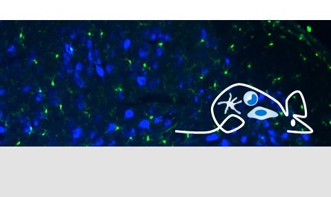
Microglia and central nervous system (CNS)-associated macrophages (CAMs), such as perivascular and meningeal macrophages, are implicated in virtually all diseases of the CNS. However, little is known about their cell-type-specific roles in the absence of suitable tools that would allow for functional discrimination between the ontogenetically closely related microglia and CAMs. To develop a new microglia gene targeting model, we first applied massively parallel single-cell analyses to compare microglia and CAM signatures during homeostasis and disease and identified hexosaminidase subunit beta (Hexb) as a stably expressed microglia core gene, whereas other microglia core genes were substantially downregulated during pathologies. Next, we generated HexbtdTomato mice to stably monitor microglia behavior in vivo. Finally, the Hexb locus was employed for tamoxifen-inducible Cre-mediated gene manipulation in microglia and for fate mapping of microglia but not CAMs. In sum, we provide valuable new genetic tools to specifically study microglia functions in the CNS.
(2020) The Journal of Clinical Investigation. 130, 3, p. 1315-1329 Abstract[All authors]Acute graft-versus-host disease (GVHD) can affect the central nervous system (CNS). The role of microglia in CNS-GVHD remains undefined. In agreement with microglia activation, we found that profound morphological changes and MHC-II and CD80 upregulation occurred upon GVHD induction. RNA sequencing-based analysis of purified microglia obtained from mice with CNS-GVHD revealed TNF upregulation. Selective TNF gene deletion in microglia of Cx3cr1creER Tnffl/- mice reduced MHC-II expression and decreased CNS T cell infiltrates and VCAM-1+ endothelial cells. GVHD increased microglia TGF-β-activated kinase-1 (TAK1) activation and NF-κB/p38 MAPK signaling. Selective Tak1 deletion in microglia using Cx3cr1creER Tak1fl/fl mice resulted in reduced TNF production and microglial MHC-II and improved neurocognitive activity. Pharmacological TAK1 inhibition reduced TNF production and MHC-II expression by microglia, Th1 and Th17 T cell infiltrates, and VCAM-1+ endothelial cells and improved neurocognitive activity, without blocking graft-versus-leukemia effects. Consistent with these findings in mice, we observed increased activation and TNF production of microglia in the CNS of GVHD patients. In summary, we prove a role for microglia in CNS-GVHD, identify the TAK1/TNF/MHC-II axis as a mediator of CNS-GVHD, and provide a TAK1 inhibitor-based approach against GVHD-induced neurotoxicity.
(2020) Oncogene. 39, 9, p. 1997-2008 Abstract[All authors]Chronic lymphocytic leukemia (CLL) is a malignancy of mature B lymphocytes. The microenvironment of the CLL cells is a vital element in the regulation of the survival of these malignant cells. CLL cell longevity is dependent on external signals, originating from cells in their microenvironment including secreted and surface-bound factors. Dendritic cells (DCs) play an important part in tumor microenvironment, but their role in the CLL bone marrow (BM) niche has not been studied. We show here that CLL cells induce accumulation of bone marrow dendritic cells (BMDCs). Depletion of this population attenuates disease expansion. Our results show that the support of the microenvironment is partly dependent on CD84, a cell surface molecule belonging to the Signaling Lymphocyte Activating Molecule (SLAM) family of immunoreceptors. Our results suggest a novel therapeutic strategy whereby eliminating BMDCs or blocking the CD84 expressed on these cells may reduce the tumor load.
(2020) European Journal of Immunology. 50, 4, p. 537-547 AbstractThe small intestine hosts specialized lymphoid structures, the Peyer's patches, that face the gut lumen and are overlaid with unique epithelial cells, called microfold (M) cells. M cells are considered to constitute an important route for antigen uptake in the mucosal immune system. Here, we used intravital microscopy to define immune cell populations, which are in close contact with M cells and potentially sample antigen. We present live evidence that DCs enter M cell pockets and highlight the abundance of mononuclear phagocytes in these structures. Taking advantage of the respective reporter animals, we focused on classical DCs that express Zbtb46 and analyzed how these cells interact with M cells in steady state and sample antigen for T cell activation in the Peyer's patches following challenge.
(2020) Frontiers in Immunology. 11, 614509. Abstract[All authors]Systemic inflammation is associated with alterations in complex brain functions such as learning and memory. However, diagnostic approaches to functionally assess and quantify inflammation-associated alterations in synaptic plasticity are not well-established. In previous work, we demonstrated that bacterial lipopolysaccharide (LPS)-induced systemic inflammation alters the ability of hippocampal neurons to express synaptic plasticity, i.e., the long-term potentiation (LTP) of excitatory neurotransmission. Here, we tested whether synaptic plasticity induced by repetitive magnetic stimulation (rMS), a non-invasive brain stimulation technique used in clinical practice, is affected by LPS-induced inflammation. Specifically, we explored brain tissue cultures to learn more about the direct effects of LPS on neural tissue, and we tested for the plasticity-restoring effects of the anti-inflammatory cytokine interleukin 10 (IL10). As shown previously, 10 Hz repetitive magnetic stimulation (rMS) of organotypic entorhino-hippocampal tissue cultures induced a robust increase in excitatory neurotransmission onto CA1 pyramidal neurons. Furthermore, LPS-treated tissue cultures did not express rMS-induced synaptic plasticity. Live-cell microscopy in tissue cultures prepared from a novel transgenic reporter mouse line [C57BL/6-Tg(TNFa-eGFP)] confirms that ex vivo LPS administration triggers microglial tumor necrosis factor alpha (TNFα) expression, which is ameliorated in the presence of IL10. Consistent with this observation, IL10 hampers the LPS-induced increase in TNFα, IL6, IL1β, and IFNγ and restores the ability of neurons to express rMS-induced synaptic plasticity in the presence of LPS. These findings establish organotypic tissue cultures as a suitable model for studying inflammation-induced alterations in synaptic plasticity, thus providing a biological basis for the diagnostic use of transcranial magnetic stimulation in the context of brain inflammation.
(2020) Immunity. 53, 5, p. 1033-1049.e7 Abstract[All authors]Microglia, the resident macrophages of the brain parenchyma, are key players in central nervous system (CNS) development, homeostasis, and disorders. Distinct brain pathologies seem associated with discrete microglia activation modules. How microglia regain quiescence following challenges remains less understood. Here, we explored the role of the interleukin-10 (IL-10) axis in restoring murine microglia homeostasis following a peripheral endotoxin challenge. Specifically, we show that lipopolysaccharide (LPS)-challenged mice harboring IL-10 receptor-deficient microglia displayed neuronal impairment and succumbed to fatal sickness. Addition of a microglial tumor necrosis factor (TNF) deficiency rescued these animals, suggesting a microglia-based circuit driving pathology. Single cell transcriptome analysis revealed various IL-10 producing immune cells in the CNS, including most prominently Ly49D+ NK cells and neutrophils, but not microglia. Collectively, we define kinetics of the microglia response to peripheral endotoxin challenge, including their activation and robust silencing, and highlight the critical role of non-microglial IL-10 in preventing deleterious microglia hyperactivation.
(2020) The EMBO Journal. 39, 22, e104464. Abstract[All authors]Microglia are the principal phagocytes that clear cell debris in the central nervous system (CNS). This raises the question, which cells remove cell debris when microglial phagocytic activity is impaired. We addressed this question using Siglechdtr mice, which enable highly specific ablation of microglia. Non-microglial mononuclear phagocytes, such as CNS-associated macrophages and circulating inflammatory monocytes, did not clear microglial debris. Instead, astrocytes were activated, exhibited a pro-inflammatory gene expression profile, and extended their processes to engulf microglial debris. This astrocytic phagocytosis was also observed in Irf8-deficient mice, in which microglia were present but dysfunctional. RNA-seq demonstrated that even in a healthy CNS, astrocytes express TAM phagocytic receptors, which were the main astrocytic phagocytic receptors for cell debris in the above experiments, indicating that astrocytes stand by in case of microglial impairment. This compensatory mechanism may be important for the maintenance or prolongation of a healthy CNS.
(2020) Nature Immunology. 21, 5, p. 525-534 Abstract[All authors]Mildner and colleagues characterize two subsets (Cxcl10(+) and Saa3(+)) of monocytes with pathogenic potential in the central nervous system of mice with experimentally induced autoimmune encephalomyelitis and show these pathogenic cells are not derived from Ly6C(+) monocytes, but from early myeloid cell progenitors.Multiple sclerosis (MS) is characterized by pathological inflammation that results from the recruitment of lymphoid and myeloid immune cells from the blood into the brain. Due to subset heterogeneity, defining the functional roles of the various cell subsets in acute and chronic stages of MS has been challenging. Here, we used index and transcriptional single-cell sorting to characterize the mononuclear phagocytes that infiltrate the central nervous system from the periphery in mice with experimentally induced autoimmune encephalomyelitis, a model of MS. We identified eight monocyte and three dendritic cell subsets at acute and chronic disease stages in which the defined transcriptional programs pointed toward distinct functions. Monocyte-specific cell ablation identified Cxcl10(+) and Saa3(+) monocytic subsets with a pathogenic potential. Transfer experiments with different monocyte and precursor subsets indicated that these Cxcl10(+) and Saa3(+) pathogenic cells were not derived from Ly6C(+) monocytes but from early myeloid cell progenitors. These results suggest that blocking specific pathogenic monocytic subsets, including Cxcl10(+) and Saa3(+) monocytes, could be used for targeted therapeutic interventions.
(2020) Journal of Immunology. 205, 10, p. 2583-2594 Abstract[All authors]Protective MHC class I-dependent immune responses require an overlap between repertoires of proteins directly presented on target cells and cross-presented by professional APC, specifically dendritic cells. How stable proteins that rely on defective ribosomal proteins for direct presentation are captured for cell-to-cell transfer remains enigmatic. In this study, we address this issue using a combination of in vitro (C57BL/6-derived mouse cell lines) and in vivo (C57BL/6 mouse strains) approaches involving stable and unstable versions of OVA model Ags displaying defective ribosomal protein-dependent and -independent Ag presentation, respectively. Apoptosis, but not necrosis, of donor cells was found associated with robust global protein aggregate formation and captured stable proteins permissive for cross-presentation. Potency of aggregates to serve as Ag source was directly demonstrated using polyglutamine-equipped model substrates. Collectively, our data implicate global protein aggregation in apoptotic cells as a mechanism that ensures the overlap between MHC class I epitopes presented directly or cross-presented by APC and demonstrate the unusual ability of dendritic cells to process stable protein aggregates.
(2020) Immunology Letters. 227, p. 66-78 AbstractMonocytes are circulating myeloid immune precursor cells that are generated in the bone marrow. Mature monocytes are released into the circulation and, in case of need, recruited to peripheral sites of inflammation to differentiate into monocyte-derived effector cells. In absence of overt inflammation, monocytes also extravasate into selected tissues, where they complement tissue-resident macrophage compartments. Adjustment of these homeostatic monocyte infiltrates to local environment is critical to maintain health, as best established for the intestine. Defined gene expression changes that differ between gut segments presumably help strike the fine balance between the crucial function of these monocyte-derived macrophages as tissue rheostats and their detrimental hyperactivation. Environmental factors that dictate local monocyte differentiation remain incompletely understood. Definition of the latter could aid our general understanding of in vivo monocyte functions and their relation to inflammatory disorders. In this review, we summarize recent advances in our understanding of monocyte subsets, their differentiation into tissue macrophages, and selected contributions of monocyte-derived cells to steady-state physiology. Moreover, we will discuss emerging evidence for an intriguing bifurcation of monocyte development in the bone marrow and potential functional implications. Emphasis will be given to points of controversies, but we will largely focus on the healthy organism. For a discussion of monocyte and macrophage contributions to inflammatory conditions, we refer the reader to other dedicated reviews.
(2020) Journal of Immunology. 204, 3, p. 707-717 Abstract[All authors]Recruited blood monocytes contribute to the establishment, perpetuation, and resolution of tissue inflammation. Specifically, in the inflamed intestine, monocyte ablation was shown to ameliorate colitis scores in preclinical animal models. However, the majority of intestinal macrophages that seed the healthy gut are also monocyte derived. Monocyte ablation aimed to curb inflammation would therefore likely interfere with intestinal homeostasis. In this study, we used a TLR2 trans-membrane peptide that blocks TLR2 dimerization that is critical for TLR2/1 and TLR2/6 heterodimer signaling to blunt inflammation in a murine colitis model. We show that although the TLR2 peptide treatment ameliorated colitis, it allowed recruited monocytes to give rise to macrophages that lack the detrimental proinflammatory gene signature and reduced potentially damaging neutrophil infiltrates. Finally, we demonstrate TLR blocking activity of the peptide on in vitro cultured human monocyte-derived macrophages. Collectively, we provide a significantly improved anti-inflammatory TLR2 peptide and critical insights in its mechanism of action toward future potential use in the clinic.
(2020) eLife. 9, e49998. Abstract[All authors]Monocytes are circulating short-lived macrophage precursors that are recruited on demand from the blood to sites of inflammation and challenge. In steady state, classical monocytes give rise to vasculature-resident cells that patrol the luminal side of the endothelium. In addition, classical monocytes feed macrophage compartments of selected organs, including barrier tissues, such as the skin and intestine, as well as the heart. Monocyte differentiation under conditions of inflammation has been studied in considerable detail. In contrast, monocyte differentiation under non-inflammatory conditions remains less well understood. Here we took advantage of a combination of cell ablation and precursor engraftment to investigate the generation of gut macrophages from monocytes. Collectively, we identify factors associated with the gradual adaptation of monocytes to tissue residency. Moreover, comparison of monocyte differentiation into the colon and ileum-resident macrophages revealed the graduated acquisition of gut segment-specific gene expression signatures.
2019
-
(2019) Blood. 134, 16, p. 1274-1275 Abstract
Hematopoiesis is well known to be affected by environmental factors, adjusting the balance of lymphoid and myeloid output according to peripheral needs.(1,2) Specifically, the bone marrow (BM), as a site of adult blood cell generation, has been shown to sense the gut microbiome composition and respond to dysbiosis associated with antibiotics treatment and numerous gastrointestinal disorders. In this issue of Blood, Lee et al(3) describe how this remote sensing is achieved and how the microbiota educate the immune system while maintaining critical steady-state myelopoiesis.(4)
(2019) Nature Nanotechnology. 14, 9, p. 891-901 Abstract[All authors]A low response rate, acquired resistance and severe side effects have limited the clinical outcomes of immune checkpoint therapy. Here, we show that combining cancer nanovaccines with an anti-PD-1 antibody (alpha PD-1) for immunosuppression blockade and an anti-OX40 antibody (alpha OX40) for effector T-cell stimulation, expansion and survival can potentiate the efficacy of melanoma therapy. Prophylactic and therapeutic combination regimens of dendritic cell-targeted mannosylated nanovaccines with alpha PD-1/alpha OX40 demonstrate a synergism that stimulates T-cell infiltration into tumours at early treatment stages. However, this treatment at the therapeutic regimen does not result in an enhanced inhibition of tumour growth compared to alpha PD-1/alpha OX40 alone and is accompanied by an increased infiltration of myeloid-derived suppressor cells in tumours. Combining the double therapy with ibrutinib, a myeloid-derived suppressor cell inhibitor, leads to a remarkable tumour remission and prolonged survival in melanoma-bearing mice. The synergy between the mannosylated nanovaccines, ibrutinib and alpha PD-1/alpha OX40 provides essential insights to devise alternative regimens to improve the efficacy of immune checkpoint modulators in solid tumours by regulating the endogenous immune response.
(2019) eLife. 8, 42025. Abstract[All authors]A characteristic subset of microglia expressing CD11c appears in response to brain damage. However, the functional role of CD11c(+) microglia, as well as the mechanism of its induction, are poorly understood. Here we report that the genetic ablation of signal regulatory protein alpha (SIRP alpha), a membrane protein, induced the emergence of CD11c(+) microglia in the brain white matter. Mice lacking CD47, a physiological ligand of SIRP alpha, and microglia-specific SIRP alpha-knockout mice exhibited the same phenotype, suggesting that an interaction between microglial SIRP alpha and CD47 on neighbouring cells suppressed the emergence of CD11c(+) microglia. A lack of SIRP alpha did not cause detectable damage to the white matter, but resulted in the increased expression of genes whose expression is characteristic of the repair phase after demyelination. In addition, cuprizone-induced demyelination was alleviated by the microglia-specific ablation of SIRP alpha. Thus, microglial SIRP alpha suppresses the induction of CD11c(+) microglia that have the potential to accelerate the repair of damaged white matter.
(2019) European Journal of Immunology. 50, 3, p. 353-362 Abstract[All authors]Conditional mutagenesis and fate mapping have contributed considerably to our understanding of physiology and pathology. Specifically, Cre recombinase-based approaches allow the definition of cell type-specific contributions to disease development and of inter-cellular communication circuits in respective animal models. Here we compared Cx(3)cr1(CreER) and Sall1(CreER) transgenic mice and their use to decipher the brain macrophage compartment as a showcase to discuss recent technological advances. Specifically, we highlight the need to define the accuracy of Cre recombinase expression, as well as strengths and pitfalls of these particular systems that should be taken into consideration when applying these models.
(2019) Cell. 179, 2, p. 292-311 AbstractMicroglia were first recognized as a distinct cell population in the CNS one century ago. For a long time, they were primarily considered to be phagocytes responsible for removing debris during CNS development and disease. More recently, advances in imaging and genetics and the advent of single-cell technology provided new insights into the much more complex and fascinatir; biology of microglia. The ontogeny of microglia was identified, and their functions in health and disease were better defined. Although many qL lions about microglia and their roles in human diseases remain unanswered, the prospect of targeting microglia for the treatment of neurological and psychiatric disorders is tantalizing.
(2019) Science immunology. 4, 36, 6571. Abstract[All authors]Cytokines maintain intestinal health, but precise intercellular communication networks remain poorly understood. Macrophages are immune sentinels of the intestinal tissue and are critical for gut homeostasis. Here, we show that in a murine inflammatory bowel disease (IBD) model based on macrophage-restricted interleukin-10 (IL-10) receptor deficiency (Cx3cr1Cre:Il10rafl/fl mice), proinflammatory mutant gut macrophages cause severe spontaneous colitis resembling the condition observed in children carrying IL-10R mutations. We establish macrophage-derived IL-23 as the driving factor of this pathology. Specifically, we report that Cx3cr1Cre:Il10rafl/fl:Il23afl/fl mice harboring macrophages deficient for both IL-10R and IL-23 are protected from colitis. By analyzing the epithelial response to proinflammatory macrophages, we provide evidence that T cells of colitic animals produce IL-22, which induces epithelial chemokine expression and detrimental neutrophil recruitment. Collectively, we define macrophage-specific contributions to the induction and pathogenesis of colitis, as manifested in mice harboring IL-10R deficiencies and human IBDs.
(2019) European Journal of Immunology. 49, 1, p. 19-29 AbstractThe involvement of macrophages in the pathogenesis of obesity has been recognized since 2003. Early studies mostly focused on the role of macrophages in adipose tissue (AT) and in obesity-associated chronic low-grade inflammation. Lately, AT macrophages were shown to undergo intrinsic metabolic changes that affect their immune function (i.e., immunometabolism), corresponding to their unique properties along the range of pro- versus anti-inflammatory activity. In parallel, recent studies in mice revealed critical neuronal-macrophage interactions, both in the CNS and in peripheral tissues, including in white and brown AT. These intercellular activities impinge on energy and metabolic homeostasis, partially by also engaging adipocytes in a neuronal-macrophage-adipocyte menage a trois. Finally, neuropeptides (NP), such as NPY and appetite-reducing NPFF, may prove as mediators in such intercellular network. In this concise review, we highlight some of these recent insights on adipose macrophage immunometabolism, as well as central and peripheral neuronal-macrophage interactions with emphasis on their impact on adipocyte biology and whole-body metabolism. We also discuss the expanding view on the role of the NP, NPY and NPFF, in obesity.
(2019) Frontiers in Immunology. 10, APR, 863. Abstract[All authors]Dendritic cells (DC) are unrivaled in their potential to prime naive T cells by presenting antigen and providing costimulation. DC are furthermore believed to decode antigen context by virtue of pattern recognition receptors and to polarize T cells through cytokine secretion toward distinct effector functions. Diverse polarized T helper (T-H) cells have been explored in great detail. In contrast, studies of instructing DC have to date largely been restricted to in vitro settings or adoptively transferred DC. Here we report efforts to unravel the DC response to cognate T cell encounter in antigen-challenged lymph nodes (LN). Mice engrafted with antigen-specific T cells were immunized with nanoparticles (NP) entrapping adjuvants and absorbed with antigen to study the immediate DC response to T cell encounter using bulk and single cell RNA-seq profiling. NP induced robust antigen-specific T(H)1 cell responses with minimal bystander activation. Fluorescent-labeled NP allowed identification of antigen-carrying DC and focus on transcriptional changes in DC that encounter T cells. Our results support the existence of a bi-directional crosstalk between DC and T cells that promotes T(H)1 responses, including involvement of the ubiquitin-like molecule Isg15 that merits further study.
2018
-
(2018) Journal of Neuroinflammation. 15, 1, 278. Abstract[All authors]
Background: Fractalkine (CX(3)CL1) and its receptor (CX(3)CR1) play an important role in regulating microglial function. We have previously shown that Cx(3)cr1 deficiency exacerbated tau pathology and led to cognitive impairment. However, it is still unclear if the chemokine domain of the ligand CX(3)CL1 is essential in regulating neuronal tau pathology.Methods: We used transgenic mice lacking endogenous Cx(3)cl1 (Cx(3)cl1(-/-)) and expressing only obligatory soluble form (with only chemokine domain) and lacking the mucin stalk of CX(3)CL1 (referred to as Cx(3)cl1(105 Delta) mice) to assess tau pathology and behavioral function in both lipopolysaccharide (LPS) and genetic (hTau) mouse models of tauopathy.Results: First, increased basal tau levels accompanied microglial activation in Cx(3)cl1(105 Delta) mice compared to control groups. Second, increased CD45(+) and F4/80(+) neuroinflammation and tau phosphorylation were observed in LPS, hTau/Cx(3)cl1(-/-), and hTau/Cx(3)cl1(105 Delta) mouse models of tau pathology, which correlated with impaired spatial learning. Finally, microglial cell surface expression of CX(3)CR1 was reduced in Cx(3)cl1(105 Delta) mice, suggesting enhanced fractalkine receptor internalization (mimicking Cx(3)cr1 deletion), which likely contributes to the elevated tau pathology.Conclusions: Collectively, our data suggest that overexpression of only chemokine domain of CX(3)CL1 does not protect against tau pathology.
(2018) Nature Immunology. 19, 6, p. 636-644 Abstract[All authors]Transcriptome profiling is widely used to infer functional states of specific cell types, as well as their responses to stimuli, to define contributions to physiology and pathophysiology. Focusing on microglia, the brain's macrophages, we report here a side-by-side comparison of classical cell-sorting-based transcriptome sequencing and the 'RiboTag' method, which avoids cell retrieval from tissue context and yields translatome sequencing information. Conventional whole-cell microglial transcriptomes were found to be significantly tainted by artifacts introduced by tissue dissociation, cargo contamination and transcripts sequestered from ribosomes. Conversely, our data highlight the added value of RiboTag profiling for assessing the lineage accuracy of Cre recombinase expression in transgenic mice. Collectively, this study indicates method-based biases, reveals observer effects and establishes RiboTag-based translatome profiling as a valuable complement to standard sorting-based profiling strategies.
(2018) Cell Reports. 23, 7, p. 1962-1976 Abstract[All authors]Nitric oxide (NO) plays an established role in numerous physiological and pathological processes, but the specific cellular sources of NO in disease pathogenesis remain unclear, preventing the implementation of NO-related therapy. Argininosuccinate lyase (ASL) is the only enzyme able to produce arginine, the substrate for NO generation by nitric oxide synthase (NOS) isoforms. Here, we generated cell-specific conditional ASL knockout mice in combination with genetic and chemical colitis models. We demonstrate that NO derived from enterocytes alleviates colitis by decreasing macrophage infiltration and tissue damage, whereas immune cell-derived NO is associated with macrophage activation, resulting in increased severity of inflammation. We find that induction of endogenous NO production by enterocytes with supplements that upregulate ASL expression and complement its substrates results in improved epithelial integrity and alleviation of colitis and of inflammation-associated colon cancer.
(2018) Nature Communications. 9, 1, 5206. Abstract[All authors]Microglia are yolk sac-derived macrophages residing in the parenchyma of brain and spinal cord, where they interact with neurons and other glial. After different conditioning paradigms and bone marrow (BM) or hematopoietic stem cell (HSC) transplantation, graft-derived cells seed the brain and persistently contribute to the parenchymal brain macrophage compartment. Here we establish that graft-derived macrophages acquire, over time, microglia characteristics, including ramified morphology, longevity, radio-resistance and clonal expansion. However, even after prolonged CNS residence, transcriptomes and chromatin accessibility landscapes of engrafted, BM-derived macrophages remain distinct from yolk sac-derived host microglia. Furthermore, engrafted BM-derived cells display discrete responses to peripheral endotoxin challenge, as compared to host microglia. In human HSC transplant recipients, engrafted cells also remain distinct from host microglia, extending our finding to clinical settings. Collectively, our data emphasize the molecular and functional heterogeneity of parenchymal brain macrophages and highlight potential clinical implications for HSC gene therapies aimed to ameliorate lysosomal storage disorders, microgliopathies or general monogenic immuno-deficiencies.
(2018) European Journal of Immunology. 48, 8, p. 1308-1318 Abstract[All authors]Microglia are resident immune cells in the CNS, strategically positioned to clear dead cells and debris, and orchestrate CNS inflammation and immune defense. In steady state, these macrophages lack MHC class II (MHCII) expression, but microglia activation can be associated with MHCII induction. Whether microglial MHCII serves antigen presentation for critical local T-cell restimulation in CNS auto-immune disorders or modulates microglial signaling output remains under debate. To probe for such scenarios, we generated mice harboring an MHCII deficiency in microglia, but not peripheral myeloid cells. Using the CX(3)CR1(CreER)-based approach we report that microglial antigen presentation is obsolete for the establishment of EAE, with disease onset, progression, and severity unaltered in mutant mice. Antigen presentation-independent roles of microglial MHCII were explored using a demyelination model induced by the copper chelator cuprizone. Absence of microglial I-A(b) did not affect the extent of these chemically induced white matter alterations, nor did it affect microglial proliferation or gene expression associated with locally restricted de- and remyelination.
(2018) Nature Communications. 9, 1, 2036. Abstract[All authors]Microglia, the mononuclear phagocytes of the central nervous system (CNS), are important for the maintenance of CNS homeostasis, but also critically contribute to CNS pathology. Here we demonstrate that the nuclear factor kappa B (NF-kappa B) regulatory protein A20 is crucial in regulating microglia activation during CNS homeostasis and pathology. In mice, deletion of A20 in microglia increases microglial cell number and affects microglial regulation of neuronal synaptic function. Administration of a sublethal dose of lipopolysaccharide induces massive microglia activation, neuroinflammation, and lethality in mice with microgliaconfined A20 deficiency. Microglia A20 deficiency also exacerbates multiple sclerosis (MS) like disease, due to hyperactivation of the NIrp3 inflammasome leading to enhanced interleukin-113 secretion and CNS inflammation. Finally, we confirm a NIrp3 inflammasome signature and IL-1 beta expression in brain and cerebrospinal fluid from MS patients. Collectively, these data reveal a critical role for A20 in the control of microglia activation and neuroinflammation.
(2018) Nanomedicine: Nanotechnology, Biology, and Medicine. 14, 3, p. 835-847 Abstract[All authors]Nanoparticulate vaccines are promising tools to overcome cancer immune evasion. However, a deeper understanding on nanoparticle-immune cell interactions and treatments regime is required for optimal efficacy. We provide a comprehensive study of treatment schedules and mode of antigen-association to nanovaccines on the modulation of T cell immunity in vivo, under steady-state and tumor-bearing mice. The coordinated delivery of antigen and two adjuvants (Monophosphoryl lipid A, oligodeoxynucleotide cytosine-phosphate-guanine motifs (CpG)) by nanoparticles was crucial for dendritic cell activation. A single vaccination dictated a 3-fold increase on cytotoxic memory-T cells and raised antigen-specific immune responses against B16.M05 melanoma. It generated at least a 5-fold increase on IFN-gamma cytokine production, and presented over 50% higher lymphocyte count in the tumor microenvironment, compared to the control. The number of lymphocytes at the tumor site doubled with triple immunization. This lymphocyte infiltration pattern was confirmed in mammary huHER2 carcinoma, with significant tumor reduction. (c) 2018 Elsevier Inc. All rights reserved.
ICAMs Are Not Obligatory for Functional Immune Synapses between Naive CD4 T Cells and Lymph Node DCs(2018) Cell Reports. 22, 4, p. 849-859 Abstract[All authors]Protective immune responses depend on the formation of immune synapses between T cells and antigen-presenting cells (APCs). The two main LFA-1 ligands, ICAM-1 and ICAM-2, are co-expressed on many cell types, including APCs and blood vessels. Although these molecules were suggested to be key players in immune synapses studied in vitro, their contribution to helper T cell priming in vivo is unclear. Here, we used transgenic mice and intravital imaging to examine the role of dendritic cell (DC) ICAM-1 and ICAM-2 in naive CD4 T cell priming and differentiation in skin-draining lymph nodes. Surprisingly, ICAM deficiency on endogenous CD40-stimulated lymph node DCs did not impair their ability to arrest and prime CD4 lymphocyte activation and differentiation into Th1 and Tfh effectors. Thus, functional T cell receptor (TCR)-specific helper T cell synapses with antigen-presenting DCs and subsequent proliferation and early differentiation into T effectors do not require LFA-1-mediated T cell adhesiveness to DC ICAMs.
(2018) European Journal of Immunology. 48, 7, p. 1114-1119 AbstractCell ablation is a valuable complement to mutagenesis for experimentally defining specific cell functions in physiology and pathophysiology in small animal models. One of the most popular ablation strategies involves transgenic expression of a primate diphtheria toxin receptor (DTR) on murine cells that are otherwise resistant to the bacterial exotoxin. The efforts of many laboratories using the DTR approach over the years have yielded numerous valuable insights into specific cell functions. Here, we will discuss the technical aspects of the DTR approach, including the strengths, pitfalls, and future strategies to overcome the shortcomings, highlighting a recent paper published in the European Journal of Immunology [El Hachem etal. Eur. J. Immunol. 2018 ]. A particular focus will be given to the application of DTR approach to decipher in vivo functions of the murine myeloid cell compartment.
2017
-
(2017) Haematologica. 102, 12, p. 476-480 Abstract[All authors]
Hematopoietic-specific miR-142 is a critical regulator of various blood cell lineages including CD4+ dendritic cells1 and platelet biogenesis in megakaryocytes.2 Furthermore, we recently reported that miR-142 is required in order to maintain the biconcave shape of erythrocytes, their structural resilience and lifespan, through a mechanism that involves actin filament homeostasis.3 Here, we used a mouse loss of function allele to characterize a new axis, where miR-142 functions upstream of Rac1 in regulating erythropoiesis.
(2017) Journal of Controlled Release. 258, p. 182-195 Abstract[All authors]Vaccination is a promising strategy to trigger and boost immune responses against cancer or infectious disease. We have designed, synthesized and characterized aliphatic-polyester (poly(lactic-co-glycolic acid) (PLGA) nanoparticles (NP) to investigate how the nature of protein association (adsorbed versus entrapped) and polymer/surfactant concentrations impact on the generation and modulation of antigen-specific immune responses. The ability of the NP formulations to target dendritic cells (DC), be internalized and activate the T cells was characterized and optimized in vitro and in vivo using markers of DC activation and co-stimulatory molecules. Ovalbumin (OVA) was used as a model antigen in combination with the engraftment of CD4+ and CD8+ T cells, carrying a transgenic OVA-responding T cell receptor (TCR), to trace and characterize the activation of antigen-specific CD4+ and CD8+ lymph node T cells upon NP vaccination. Accordingly, the phenotype and frequency of immune cell stimulation induced by the NP loaded with OVA, isolated or in combination with synthetic unmethylated cytosine-phosphate-guanine (CpG) oligodeoxynucleotide (ODN) motifs, were characterized. DC-NP interactions increased with incubation time, presenting internalization values between 50 and 60% and 3040%, in vitro and in vivo, respectively. Interestingly, animal immunization with antigen-adsorbed NP up-regulated major histocompatibility complex (MHC) class II (MHCII), while NP entrapping the antigen up-regulated MHCI, suggesting a more efficient cross-presentation. On the other hand, rather surprisingly, the surfactant used in the NP formulation had a major impact on the activation of antigen presenting cells (APC). In fact, DC collected from lymph nodes of animals immunized with NP prepared using poly(vinil alcohol) (PVA), as a surfactant, expressed significantly higher levels of CD86, MHCI and MHCII. In addition, those NP prepared with PVA and co-entrapping OVA and the toll-like receptor (TLR) ligand CpG, induced the most profound antigen-specific T cell response, by both CD4+ and CD8+ T cells, in vivo. Overall, our data reveal the impact of NP composition and surface properties on the type and extension of induced immune responses. Deeper understanding on the NP-immune cell crosstalk can guide the rational development of nano-immunotherapeutic systems with improved and specific therapeutic efficacy and avoiding off-target effects.
(2017) Immunity. 47, 1, p. 183-198.e6 Abstract[All authors]Tissue macrophages arise during embryogenesis from yolk-sac (YS) progenitors that give rise to primitive YS macrophages. Until recently, it has been impossible to isolate or derive sufficient numbers of YS-derived macrophages for further study, but data now suggest that induced pluripotent stem cells (iPSCs) can be driven to undergo a process reminiscent of YS-hematopoiesis in vitro. We asked whether iPSC-derived primitive macrophages (iMacs) can terminally differentiate into specialized macrophages with the help of growth factors and organ-specific cues. Co-culturing human or murine iMacs with iPSC-derived neurons promoted differentiation into microglia-like cells in vitro. Furthermore, murine iMacs differentiated in vivo into microglia after injection into the brain and into functional alveolar macrophages after engraftment in the lung. Finally, iPSCs from a patient with familial Mediterranean fever differentiated into iMacs with pro-inflammatory characteristics, mimicking the disease phenotype. Altogether, iMacs constitute a source of tissue-resident macrophage precursors that can be used for biological, pathophysiological, and therapeutic studies. Yolk-sac (YS) embryonic macrophages contribute to tissue-resident macrophages but remain difficult to study because of their stage-dependent limited availability. Takata et al. demonstrate that iPSCs can generate YS macrophage-like cells (iMacs) that differentiate into functional tissue-resident macrophage-like cells upon receiving organ-specific cues, thus providing a platform for modeling tissue-resident macrophages.
(2017) Frontiers in Immunology. 8, JUN, 626. Abstract[All authors]Monocyte-derived macrophages (MoMF) play a pivotal role in the resolution of acetaminophen-induced liver injury (AILI). Timely termination of neutrophil activity and their clearance are essential for liver regeneration following injury. Here, we show that infiltrating Ly6Chi monocytes, their macrophage descendants, and neutrophils spatially and temporally overlap in the centrilobular necrotic areas during the necroinflammatory and resolution phases of AILI. At the necroinflammatory phase, inducible ablation of circulating Ly6Chi monocytes resulted in reduced numbers and fractions of reactive oxygen species (ROS)-producing neutrophils. In alignment with this, neutrophils sorted from monocyte-deficient livers exhibited reduced expression of NADPH oxidase 2. Moreover, human CD14+ monocytes stimulated with lipopolysaccharide or hepatocyte apoptotic bodies directly induced ROS production by cocultured neutrophils. RNA-seq-based transcriptome profiling of neutrophils from Ly6Chi monocyte-deficient versus normal livers revealed 449 genes that were differentially expressed with at least twofold change (p ≤ 0.05). In the absence of Ly6Chi monocytes, neutrophils displayed gene expression alterations associated with decreased innate immune activity and increased cell survival. At the early resolution phase, Ly6Chi monocytes differentiated into ephemeral Ly6Clo MoMF and their absence resulted in significant accumulation of late apoptotic neutrophils. Further gene expression analysis revealed the induced expression of a specific repertoire of bridging molecules and receptors involved with apoptotic cell clearance during the transition from Ly6Chi monocytes to MoMF. Collectively, our findings establish a phase-dependent task division between liver-infiltrating Ly6Chi monocytes and their MoMF descendants with the former regulating innate immune functions and cell survival of neutrophils and the later neutrophil clearance.
(2017) Immunity. 46, 6, p. 1030-1044.e8 Abstract[All authors]Microglia seed the embryonic neuro-epithelium, expand and actively sculpt neuronal circuits in the developing central nervous system, but eventually adopt relative quiescence and ramified morphology in the adult. Here, we probed the impact of post-transcriptional control by microRNAs (miRNAs) on microglial performance during development and adulthood by generating mice lacking microglial Dicer expression at these distinct stages. Conditional Dicer ablation in adult microglia revealed that miRNAs were required to limit microglial responses to challenge. After peripheral endotoxin exposure, Dicer-deficient microglia expressed more pro-inflammatory cytokines than wild-type microglia and thereby compromised hippocampal neuronal functions. In contrast, prenatal Dicer ablation resulted in spontaneous microglia activation and revealed a role for Dicer in DNA repair and preservation of genome integrity. Accordingly, Dicer deficiency rendered otherwise radio-resistant microglia sensitive to gamma irradiation. Collectively, the differential impact of the Dicer ablation on microglia of the developing and adult brain highlights the changes these cells undergo with time. Microglia of developing and adult brain differ in activation state and function. Here, Varol and colleagues ablated microglial Dicer expression at distinct times. Adult microglia tolerated the perturbation but became hyper-responsive to challenge compromising hippocampus functions. Dicer and microRNA absence during development caused spontaneous microglia activation and impaired genome integrity.
(2017) Myeloid Cells in Health and Disease. Gordon S.(eds.). p. 141-153 AbstractMonocytes are a conserved population of leukocytes that are present in all vertebrates, with some evidence of a parallel cell population in fly hemolymph (1). Monocytes are defined by their location in the bloodstream, their phenotype and nuclear morphology, as well as by their characteristic gene and microRNA expression signatures (2-5). In mice, monocytes represent 4% of the nucleated cells in the blood, with considerable marginal pools in the spleen and lungs that can be mobilized on demand (6,7). Within the blood, monocytes, and in particular the classical Ly6C+mouse subset, exhibit a characteristically short half-life of 20 h (8), akin to that of similar ephemer neutrophils (9).
(2017) Gut. 66, 12, p. 2110-2120 Abstract[All authors]Objective Postoperative ileus (POI), the most frequent complication after intestinal surgery, depends on dendritic cells (DCs) and macrophages. Here, we have investigated the mechanism that activates these cells and the contribution of the intestinal microbiota for POI induction.Design POI was induced by manipulating the intestine of mice, which selectively lack DCs, monocytes or macrophages. The disease severity in the small and large intestine was analysed by determining the distribution of orally applied fluorescein isothiocyanate-dextran and by measuring the excretion time of a retrogradely inserted glass ball. The impact of the microbiota on intestinal peristalsis was evaluated after oral antibiotic treatment.Results We found that Cd11c-Cre(+) Irf4(flox/flox) mice lack CD103(+) CD11b(+) DCs, a DC subset unique to the intestine whose function is poorly understood. Their absence in the intestinal muscularis reduced pathogenic inducible nitric oxide synthase (iNOS) production by monocytes and macrophages and ameliorated POI. Pathogenic iNOS was produced in the jejunum by resident Ly6C(-) macrophages and infiltrating chemokine receptor 2-dependent Ly6C(+) monocytes, but in the colon only by the latter demonstrating differential tolerance mechanisms along the intestinal tract. Consistently, depletion of both cell subsets reduced small intestinal POI, whereas the depletion of Ly6C(+) monocytes alone was sufficient to prevent large intestinal POI. The differential role of monocytes and macrophages in small and large intestinal POI suggested a potential role of the intestinal microbiota. Indeed, antibiotic treatment reduced iNOS levels and ameliorated POI.Conclusions Our findings reveal that CD103(+) CD11b(+) DCs and the intestinal microbiome are a prerequisite for the activation of intestinal monocytes and macrophages and for dysregulating intestinal motility in POI.
(2017) Nature Neuroscience. 20, 6, p. 793-803 Abstract[All authors]Microglia constitute a highly specialized network of tissue-resident immune cells that is important for the control of tissue homeostasis and the resolution of diseases of the CNS. Little is known about how their spatial distribution is established and maintained in vivo. Here we establish a new multicolor fluorescence fate mapping system to monitor microglial dynamics during steady state and disease. Our findings suggest that microglia establish a dense network with regional differences, and the high regional turnover rates found challenge the universal concept of microglial longevity. Microglial self-renewal under steady state conditions constitutes a stochastic process. During pathology this randomness shifts to selected clonal microglial expansion. In the resolution phase, excess disease-associated microglia are removed by a dual mechanism of cell egress and apoptosis to re-establish the stable microglial network. This study unravels the dynamic yet discrete self-organization of mature microglia in the healthy and diseased CNS.
(2017) Immunity. 46, 5, p. 849-862 Abstract[All authors]Monocytes are circulating, short-lived mononuclear phagocytes, which in mice and man comprise two main subpopulations. Murine Ly6C+ monocytes display developmental plasticity and are recruited to complement tissue-resident macrophages and dendritic cells on demand. Murine vascular Ly6C monocytes patrol the endothelium, act as scavengers, and support vessel wall repair. Here we characterized population and single cell transcriptomes, as well as enhancer and promoter landscapes of the murine monocyte compartment. Single cell RNA-seq and transplantation experiments confirmed homeostatic default differentiation of Ly6C+ into Ly6C monocytes. The main two subsets were homogeneous, but linked by a more heterogeneous differentiation intermediate. We show that monocyte differentiation occurred through de novo enhancer establishment and activation of pre-established (poised) enhancers. Generation of Ly6C monocytes involved induction of the transcription factor C/EBPb and C/EBPb-deficient mice lacked Ly6C monocytes. Mechanistically, C/EBPb bound the Nr4a1 promoter and controlled expression of this established monocyte survival factor.
(2017) Nature Immunology. 18, 6, p. 665-674 Abstract[All authors]Tissue macrophages provide immunological defense and contribute to the establishment and maintenance of tissue homeostasis. Here we used constitutive and inducible mutagenesis to delete the nuclear transcription regulator Mecp2 in macrophages. Mice that lacked the gene encoding Mecp2, which is associated with Rett syndrome, in macrophages did not show signs of neurodevelopmental disorder but displayed spontaneous obesity, which was linked to impaired function of brown adipose tissue (BAT). Specifically, mutagenesis of a BAT-resident Cx 3 Cr1 + macrophage subpopulation compromised homeostatic thermogenesis but not acute, cold-induced thermogenesis. Mechanistically, malfunction of BAT in pre-obese mice with mutant macrophages was associated with diminished sympathetic innervation and local titers of norepinephrine, which resulted in lower expression of thermogenic factors by adipocytes. Mutant macrophages overexpressed the signaling receptor and ligand PlexinA4, which might contribute to the phenotype by repulsion of sympathetic axons expressing the transmembrane semaphorin Sema6A. Collectively, we report a previously unappreciated homeostatic role for macrophages in the control of tissue innervation. Disruption of this circuit in BAT resulted in metabolic imbalance.
(2017) Nature Medicine. 23, 5, p. 623-630 Abstract[All authors]Adaptive thermogenesis is the process of heat generation in response to cold stimulation. It is under the control of the sympathetic nervous system, whose chief effector is the catecholamine norepinephrine (NE). NE enhances thermogenesis through beta 3-adrenergic receptors to activate brown adipose tissue and by 'browning' white adipose tissue. Recent studies have reported that alternative activation of macrophages in response to interleukin (IL)-4 stimulation induces the expression of tyrosine hydroxylase (TH), a key enzyme in the catecholamine synthesis pathway, and that this activation provides an alternative source of locally produced catecholamines during the thermogenic process. Here we report that the deletion of Th in hematopoietic cells of adult mice neither alters energy expenditure upon cold exposure nor reduces browning in inguinal adipose tissue. Bone marrow-derived macrophages did not release NE in response to stimulation with IL-4, and conditioned media from IL-4-stimulated macrophages failed to induce expression of thermogenic genes, such as uncoupling protein 1 (Ucp1), in adipocytes cultured with the conditioned media. Furthermore, chronic treatment with IL-4 failed to increase energy expenditure in wild-type, Ucp1(-/-) and interleukin-4 receptor-alpha double-negative (Il4ra(-/-)) mice. In agreement with these findings, adipose-tissue-resident macrophages did not express TH. Thus, we conclude that alternatively activated macrophages do not synthesize relevant amounts of catecholamines, and hence, are not likely to have a direct role in adipocyte metabolism or adaptive thermogenesis.
(2017) Haematologica. 102, 4, p. 676-685 Abstract[All authors]Hematopoietic-specific microRNA-142 is a critical regulator of various blood cell lineages, but its role in erythrocytes is unexplored. Herein, we characterize the impact of microRNA-142 on erythrocyte physiology and molecular cell biology, using a mouse loss-of-function allele. We report that microRNA-142 is required for maintaining the typical erythrocyte biconcave shape and structural resilience, for the normal metabolism of reactive oxygen species, and for overall lifespan. microRNA-142 further controls ACTIN filament homeostasis and membrane skeleton organization. The analyses presented reveal previously unappreciated functions of microRNA-142 and contribute to an emerging view of small RNAs as key players in erythropoiesis. Finally, the work herein demonstrates how a housekeeping network of cytoskeletal regulators can be reshaped by a single micro-RNA denominator in a cell type specific manner.
(2017) European Journal of Immunology. 47, 7, p. 1142-1152 Abstract[All authors]T-cell development is a spatially and temporally regulated process, orchestrated by well-defined contributions of transcription factors and cytokines. Here, we identify the noncoding RNA miR-142 as an additional regulatory layer within murine thymocyte development and proliferation. MiR-142 deficiency impairs the expression of cell cycle-promoting genes in mature mouse thymocytes and early progenitors, accompanied with increased levels of cyclin-dependent kinase inhibitor 1B (Cdkn1b, also known as p27Kip1). By using CRISPR/Cas9 technology to delete the miR-142-3p recognition element in the 3UTR of cdkn1b, we confirm that this gene is a novel target of miR-142-3p in vivo. Increased Cdkn1b protein expression alone however was insufficient to cause proliferation defects in thymocytes, indicating the existence of additional critical miR-142 targets. Collectively, we establish a key role for miR-142 in the control of early and mature thymocyte proliferation, demonstrating the multifaceted role of a single miRNA on several target genes.
(2017) Journal of Experimental Medicine. 214, 4, p. 905-917 Abstract[All authors]Monocytes are circulating mononuclear phagocytes, poised to extravasate to sites of inflammation and differentiate into monocyte-derived macrophages and dendritic cells. Tumor necrosis factor (TNF) and its receptors are up-regulated during monopoiesis and expressed by circulating monocytes, as well as effector monocytes infiltrating certain sites of inflammation, such as the spinal cord, during experimental autoimmune encephalomyelitis (EAE). In this study, using competitive in vitro and in vivo assays, we show that monocytes deficient for TNF or TNF receptors are outcompeted by their wild-type counterpart. Moreover, monocyte-autonomous TNF is critical for the function of these cells, as TNF ablation in monocytes/macrophages, but not in microglia, delayed the onset of EAE in challenged animals and was associated with reduced acute spinal cord infiltration of Ly6Chi effector monocytes. Collectively, our data reveal a previously unappreciated critical cell-autonomous role of TNF on monocytes for their survival, maintenance, and function.
2016
-
(2016) Nature Neuroscience. 19, 8, p. 995-998 Abstract[All authors]
Myelin is synthesized as a multilamellar membrane, but the mechanisms of membrane turnover are unknown. We found that myelin pieces were gradually released from aging myelin sheaths and were subsequently cleared by microglia. Myelin fragmentation increased with age and led to the formation of insoluble, lipofuscin-like lysosomal inclusions in microglia. Thus, age-related myelin fragmentation is substantial, leading to lysosomal storage and contributing to microglial senescence and immune dysfunction in aging.
(2016) International Journal of Cardiology. 209, p. 296-306 Abstract[All authors]Background The developmental origin of the c-kit expressing progenitor cell pool in the adult heart has remained elusive. Recently, it has been discovered that the injured heart is enriched with c-kit+ cells, which also express the hematopoietic marker CD45. Methods and results In this study, we characterize the phenotype and transcriptome of the c-kit +/CD45 +/CD11b +/Flk-1 +/Sca-1 ± (B-type) cell population, originating from the left atrial appendage. These cells are defined as cardiac macrophage progenitors. We also demonstrate that the CD45 + progenitor cell population activates heart development, neural crest and pluripotency-associated pathways in vitro, in conjunction with CD45 down-regulation, and acquire a c-kit +/CD45 -/CD11b -/Flk-1 -/Sca-1 + (A-type) phenotype through cell fusion and asymmetric division. This putative spontaneous reprogramming evolves into a highly proliferative, partially myogenic phenotype (C-type). Conclusions Our data suggests that A-type cells and cardiac macrophage precursor cells (B-type) have a common lineage origin, possibly resolving some current conundrums in the field of cardiac regeneration.
(2016) Nature Immunology. 17, 1, p. 18-25 AbstractMacrophages provide a critical systemic network cells of the innate immune system. Emerging data suggest that in addition, they have important tissue-specific functions that range from clearance of surfactant from the lungs to neuronal pruning and establishment of gut homeostasis. The differentiation and tissue-specific activation of macrophages require precise regulation of gene expression, a process governed by epigenetic mechanisms such as DNA methylation, histone modification and chromatin structure. We argue that epigenetic regulation of macrophages is determined by lineage- and tissue-specific transcription factors controlled by the built-in programming of myeloid development in combination with signaling from the tissue environment. Perturbation of epigenetic mechanisms of tissue macrophage identity can affect normal macrophage tissue function and contribute to pathologies ranging from obesity and autoimmunity to neurodegenerative diseases.
(2016) EMBO Journal. 35, 1, p. 89-101 Abstract[All authors]Multiple sclerosis is the most frequent chronic inflammatory disease of the CNS. The entry and survival of pathogenic T cells in the CNS are crucial for the initiation and persistence of autoimmune neuroinflammation. In this respect, contradictory evidence exists on the role of the most potent type of antigen-presenting cells, dendritic cells. Applying intravital two-photon microscopy, we demonstrate the gatekeeper function of CNS professional antigen-presenting CD11c+ cells, which preferentially interact with Th17 cells. IL-17 expression correlates with expression of GM-CSF by T cells and with accumulation of CNS CD11c+ cells. These CD11c+ cells are organized in perivascular clusters, targeted by T cells, and strongly express the inflammatory chemokines Ccl5, Cxcl9, and Cxcl10. Our findings demonstrate a fundamental role of CNS CD11c+ cells in the attraction of pathogenic T cells into and their survival within the CNS.
(2016) eLife. 5, 2016JULY, e15224. AbstractMicroglia, the resident CNS macrophages, have been implicated in the pathogenesis of Rett Syndrome (RTT), an X-linked neurodevelopmental disorder. However, the mechanism by which microglia contribute to the disorder is unclear and recent data suggest that microglia do not play a causative role. Here, we use the retinogeniculate system to determine if and how microglia contribute to pathogenesis in a RTT mouse model, the Mecp2 null mouse (Mecp2tm1.1Bird/y). We demonstrate that microglia contribute to pathogenesis by excessively engulfing, thereby eliminating, presynaptic inputs at end stages of disease (≥P56 Mecp2 null mice) concomitant with synapse loss. Furthermore, loss or gain of Mecp2 expression specifically in microglia (Cx3cr1CreER; Mecp2fl/yor Cx3cr1CreER; Mecp2LSL/y) had little effect on excessive engulfment, synapse loss, or phenotypic abnormalities. Taken together, our data suggest that microglia contribute to end stages of disease by dismantling neural circuits rendered vulnerable by loss of Mecp2 in other CNS cell types.
(2016) Nature Immunology. 17, 7, p. 797-805 Abstract[All authors]Perivascular, subdural meningeal and choroid plexus macrophages are non-parenchymal macrophages that mediate immune responses at brain boundaries. Although the origin of parenchymal microglia has recently been elucidated, much less is known about the precursors, the underlying transcriptional program and the dynamics of the other macrophages in the central nervous system (CNS). It was assumed that they have a high turnover from blood-borne monocytes. However, using parabiosis and fate-mapping approaches in mice, we found that CNS macrophages arose from hematopoietic precursors during embryonic development and established stable populations, with the notable exception of choroid plexus macrophages, which had dual origins and a shorter life span. The generation of CNS macrophages relied on the transcription factor PU.1, whereas the MYB, BATF3 and NR4A1 transcription factors were not required.
(2016) Nature Medicine. 22, 6, p. 586-597 Abstract[All authors]Astrocytes have important roles in the central nervous system (CNS) during health and disease. Through genome-wide analyses we detected a transcriptional response to type I interferons (IFN-Is) in astrocytes during experimental CNS autoimmunity and also in CNS lesions from patients with multiple sclerosis (MS). IFN-I signaling in astrocytes reduces inflammation and experimental autoimmune encephalomyelitis (EAE) disease scores via the ligand-Activated transcription factor aryl hydrocarbon receptor (AHR) and the suppressor of cytokine signaling 2 (SOCS2). The anti-inflammatory effects of nasally administered interferon (IFN)-β are partly mediated by AHR. Dietary tryptophan is metabolized by the gut microbiota into AHR agonists that have an effect on astrocytes to limit CNS inflammation. EAE scores were increased following ampicillin treatment during the recovery phase, and CNS inflammation was reduced in antibiotic-Treated mice by supplementation with the tryptophan metabolites indole, indoxyl-3-sulfate, indole-3-propionic acid and indole-3-Aldehyde, or the bacterial enzyme tryptophanase. In individuals with MS, the circulating levels of AHR agonists were decreased. These findings suggest that IFN-Is produced in the CNS function in combination with metabolites derived from dietary tryptophan by the gut flora to activate AHR signaling in astrocytes and suppress CNS inflammation.
(2016) Acta neuropathologica communications. 4, 1, 102. Abstract[All authors]Homo and heterozygote cx3cr1 mutant mice, which harbor a green fluorescent protein (EGFP) in their cx3cr1 loci, represent a widely used animal model to study microglia and peripheral myeloid cells. Here we report that microglia in the dentate gyrus (DG) of cx3cr1-/- mice displayed elevated microglial sirtuin 1 (SIRT1) expression levels and nuclear factor kappa-light-chain-enhancer of activated B cells (NF-kB) p65 activation, despite unaltered morphology when compared to cx3cr1+/- or cx3cr1+/+ controls. This phenotype was restricted to the DG and accompanied by reduced adult neurogenesis in cx3cr1-/- mice. Remarkably, adult neurogenesis was not affected by the lack of the CX3CR1-ligand, fractalkine (CX3CL1). Mechanistically, pharmacological activation of SIRT1 improved adult neurogenesis in the DG together with an enhanced performance of cx3cr1-/- mice in a hippocampusdependent learning and memory task. The reverse condition was induced when SIRT1 was inhibited in cx3cr1-/- mice, causing reduced adult neurogenesis and lowered hippocampal cognitive abilities. In conclusion, our data indicate that deletion of CX3CR1 from microglia under resting conditions modifies brain areas with elevated cellular turnover independent of CX3CL1.
(2016) Microbiology spectrum. 4, 5, MCHD-0033-. AbstractMonocytes are short-lived mononuclear phagocytes that circulate in the bloodstream and comprise two main subpopulations that in the mouse are best defined by the Ly6C marker. Intravascular functions of "classical" Ly6C+ monocytes and their interactions with other lymphoid and myeloid leukocytes in the circulation remain poorly understood. Rather, these cells are known to efficiently extravasate into tissues. Indeed, Ly6C+ monocytes and their descendants have emerged as a third, highly plastic and dynamic cellular system that complements the two classical, tissue-resident mononuclear phagocyte compartments, i.e., macrophages and dendritic cells, on demand. Following recruitment to injured tissue, Ly6C+ monocytes respond to local cues and can critically contribute to the initiation and resolution of inflammatory reactions. The second main murine monocyte subset, Ly6C- cells, derive in steady state from Ly6C+ monocytes and remain in the vasculature, where the cells act as scavengers. Moreover, a major fraction of Ly6C- monocytes adheres to the capillary endothelium and patrols the vessel wall for surveillance. Given the central role of monocytes in homeostasis and pathology, in-depth study of this cellular compartment can be highly informative on the health state of the organism and provides an attractive target for therapeutic intervention.