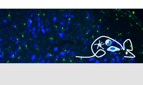
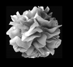
Brain macrophages comprise microglia and CNS border associated macrophages (BAM). Microglia are brain resident macrophages, which arise in the embryo, seed the developing brain and expand to an evenly distributed network in the CNS. Microglial cells subsequently maintain themselves throughout adult live by longevity and limited self-renewal. Specific functions of microglia remain incompletely understood. Emerging microglia activities include critical contributions to brain development and homeostasis by synaptic pruning and debris clearance, as well as acute contributions to neurodegeneration and – inflammation. Based on the unique high expression of the CX3CR1 chemokine receptor in microglia, we developed an experimental system that allows probing of microglia functions in their physiological context. Upon injury, the brain recruits monocytes that differentiate locally into monocyte-derived brain macrophages. These cells can functionally complement the microglia compartment. However, following physiological challenges, these cells seed the brain only transiently until a steady state, relying largely on microglia, is restored. Interestingly, monocyte-derived macrophages take with aging hold in the brain parenchyma and adopt microglia identity. Unlike microglia, monocyte-derived microglia are targets of clonal hematopoiesis and hence could contribute to brain pathology.
More recently we developped a novel binary split Cre transgenic system to discriminate functional contributions of microglia and CNS border-associated macrophages (BAM). Using this model we study the role of Lyve1 + perivascular macrophages in autoimmune neuro-degeneration (EAE) and other pathologies.
Past and present Jung lab members involved in these studies:
Alexander Mildner, Yochai Wolf, Ki-Wook Kim, Anat Shemer, Diana Varol, Alon Volasky, Neta Barashi, Zhana Haimon, Yuan Xia, Jung-Seok Kim, Jonathan Grozovski, Liron Levy, Gal Frumer, Sun Hye Shin, Lior Schori.
Publications:
Jung et al. MCB 2000
Shechter, Varol et al. London et al. PLOS Medicine, 2009
Wolf et al. Front. in Cell. Neuroscience 2013 (review)
Goldmann et al. Nat Neuroscience 2013
Lavin et al. Cell 2014
Shemer and Jung. Semin. Immunopath 2015 (review)
Bruttger et al. Immunity 2015 (collaboration with Waisman lab)
Shemer et al. Trend in Immunology 2015 (review)
Varol et al. Immunity 2017
Haimon et al. Nat Immunol 2018
Wolf, Shemer, Levy-Efrati et al. Eur J Immunol 2018
Shemer et al. Nat Comm, 2018;
Prinz, Jung and Priller. Cell 2019 (review)
Chappell-Maor et al. EJI 2019
Shemer et al. Immunity 2020
Kim et al. Immunity 2021
Paolicelli, Stevens, Sierra, Tremblay et al. Neuron 2022 (review)
Haimon et al. Nat Immunol 2021
De Schepper et al. Nat Neuroscience 2023 (collaboration with Song lab, UCL)
Frumer, Shin, Jung and Kim. Glia 2023 (review)
Kim et al. Methods and Protocols, Methods in Molecular Biology,(protocol)
Boura-Halfon et al. Methods in Molecular Biology, 2023 (protocol)
Kim et al. (bioRx)
Past and Current Funding:
DFG (FOR 1336-2, NEURO-MAC, SPP2395), ISF, MINERVA foundation, PMSA, ERA-NET Neuro
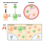
Lung Macrophages comprise cells residing in the alveolar space (AM) and cells in the interstitium (IM), that collectively play critical roles in the maintenance of pulmonary homeostasis, as well as the defense against bacterial and viral pathogen encounter. In depth understanding of differential functional contributions of these cells, as well as their origins, could pave the way for the development of novel therapeutic strategies. Using respective mouse models, we could f.i. recently show that reported lung IM subpopulations derive from distinct GMP - and MDP-derived classical monocytes. Moreover, we identified a unique macrophage population in the pulmonary visceral pleura that critically contributes to lung immune homeostasis.
Past and present Jung lab members involved in these studies:
Niss Larrossi, Ayala Raanan, Sebastien Trzebanski, Nitzan Goldmann
Publications:
Trzebanski et al., Immunity, 2024
Larrossi et al. (in preparation)
Past and Current Funding:
ISF
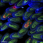
Intestinal Macrophages are abundantly dispersed throughout the lamina propria and strategically positioned key components of the gut. Unlike most tissue macrophages, these cells derive from monocytes probably recruited by a tonic low-grade inflammatory stimulus imposed by the gut microbiota. Upon arrival in healthy gut tissue monocytes are rapidly conditioned towards a non-inflammatory fate that is critical for the maintenance of gut homeostasis. Conversely, monocyte-derived cells that fail to be ‘educated’ actively perpetuate the disease state. Understanding the molecular cues that guide monocyte differentiation in small and large intestine, and their exploitation for rational monocyte manipulation might bear potential for the treatment of inflammatory bowel disorders (IBD). In the past we have given a particular focus to the role of the cytokine IL-10 in maintaining gut health.
Past and present Jung lab members involved in these studies:
Biana Bernshtein, Ehud Zigmond, Alexandra Vallon-Eberhard, Chen Varol, Tegest Aychek, Mor Gross-Vered, Caterina Curato, Masha Kolesnikov, Jung-Seok Kim, Serkalem Ayanaw, Sebastien Trzebanski, Jarmila Kralova, Anmol Kulkarni, Daichi Nonaka.
Publications:
Varol et al. JExMed 2007
Varol et al. Immunity 2009
Varol, Zigmond & Jung, Nat Rev Immunol. 2009 (review)
Zigmond et al. Immunity 2012
Zigmond et al. Trend in Immunol. 2013 (review)
Zigmond et al. Immunity 2014
Lavin et al Cell 2014
Aychek et al. Nat Communications 2015
Varol, Mildner and Jung. Annual Rev Immunol. 2015 (review)
Gross et al. Frontiers in Immunology, 2015 (review)
Shemer et al. Nat Communication 2018
Bernshtein et al. Science Immunology 2019
Kolesnikov et al. EJI 2019
Gross-Vered et al. J.Immunology 2019
Gross-Vered et al. eLife 2020
Kulkarni and Jung, Cell Metabolism, 2021, preview
Past and Current Funding:
CCFA, BSF, AdvERC
Brown Adipose Tissue Macrophages reside in the key organ responsible for homeostatic thermogenesis. We recently used constitutive and inducible mutagenesis to delete the nuclear transcription regulator methyl-CpG binding protein 2 (MeCP2) in defined tissue macrophages. Animals lacking the Rett syndrome-associated gene in macrophages did not show signs of a neurodevelopmental disorder, but surprisingly displayed altered body composition and spontaneous obesity. This phenotype involved neither hyperphagia, primary hyper-insulinemia nor inflammation, but rather could be linked to impaired brown adipose tissue (BAT) function. Specifically, mutagenesis of a BAT-resident CX3CR1+ macrophage subpopulation compromised homeostatic, though not acute cold-induced thermogenesis. Mechanistically, steady-state BAT malfunction of pre-obese mice was associated with decreased sympathetic innervation and lower local norepinephrine titers, resulting in reduced adipocyte expression of thermogenic factors. Mutant macrophages over-expressed PlexinA4, which might contribute to the phenotype by repulsion of Sema6A-expressing sympathetic axons. Collectively, we reported a previously unappreciated homeostatic role of macrophages in the control of tissue innervation, disruption of which in BAT results in a metabolic imbalance. To functionally test the role of PlexinA4 we have generated mice harboring conditional PlexinA4 alleles.
Past and present Jung lab members involved in these studies:
Yochai Wolf, Sigalit Boura-Halfon
Publications:
Wolf et al. Nat Immunology 2017
Fischer et al. Nat Medicine 2017 (collaborative study)
Boura-Halfon et al. Eur J Imm. 2019 (review)
Past and Current Funding:
ISF, AdvERC