
SUSHI: Sparsity-Based Ultrasound Super-Resolution Hemodynamic Imaging
A. Bar-Zion, O. Solomon, C. Tremblay-Darveau, D. Adam and Y. C. Eldar
Introduction
Identifying and visualizing vasculature within organs and tumors has major implications in managing cardiovascular diseases and cancer. Contrast-enhanced ultrasound scans detect slow-flowing blood, facilitating noninvasive perfusion measurements. However, their limited spatial resolution prevents the depiction of microvascular structures. Recently, superlocalization ultrasonography techniques have surpassed this limit. However, they require long acquisition times of several minutes, preventing the detection of hemodynamic changes. We present a fast super-resolution method that exploits sparsity in the underlying vasculature and statistical independence within the measured signals. Similar to super-localization techniques, this approach improves the spatial resolution by up to an order of magnitude compared to standard scans. Unlike super-localization methods, it requires acquisition times of only tens of milliseconds. We demonstrate a temporal resolution of ~25 Hz, which may enable functional super-resolution imaging deep within the tissue, surpassing the temporal resolution limitations of current superresolution methods, e.g., in neural imaging. The subsecond acquisitions make our approach robust to motion artifacts, simplifying in vivo use of super-resolution ultrasound.
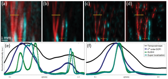
In the figure below we show the results of SUSHI in comparison to other methods. Blood vessel bifurcations, appearing in a scan acquired with high concentration of contrast agents, are depicted as (a) temporal mean, using 150 frames, (b) fourth-order SOFI, (c) SUSHI, and (d) super-localization. Red and cyan depict hemodynamic flow from and toward the transducer, respectively. Comparing visually, the super-localization image seems very noisy and unclear, while temporal mean and SOFI images have degraded resolution compared with SUSHI. SUSHI exhibits the clearest image of the vasculature, presenting a sharp, almost noiseless reconstruction, compared with the other methods. (e) and (f) Intensity profiles measured along the solid and dashed yellow lines on (a)–(d). All profiles were taken with respect to the red blood vessels only. (e) shows that in high density areas (e.g., bifurcations), SUSHI is superior, while in low density areas [e.g., isolated vessel in (f)], SUSHI exhibits comparable spatial resolution to super-localization. More details can be found in the paper below.
References
- A. Bar-Zion, O. Solomon, C. Tremblay-Darveau, D. Adam and Y. C. Eldar, "SUSHI: Sparsity-based Ultrasound Super-resolution Hemodynamic Imaging", IEEE Transactions on Ultrasonics, Ferroelectrics and Frequency Control, vol. 65, issue 12, pp. 2365-2380, December 2018.