Research

Multiplexed imaging

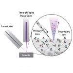
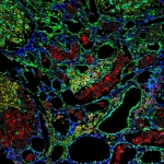
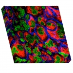
Multiplexed Ion Beam Imaging by Time of Flight (MIBI-TOF) is a new technology that enables high-dimensional single-cell imaging in situ of clinical human specimens. In MIBI-TOF, we stain human tissue sections with dozens of metal-labeled antibodies and read out their abundance and location by secondary ionization mass spectrometry. The result is a multi-dimensional image, depicting sub-cellular expression and localization for dozens of distinct proteins in situ.

Computational Tools for Image Analysis

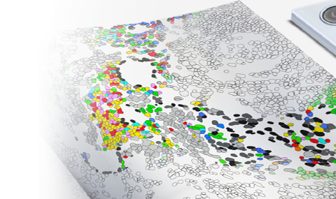
Our lab develops state-of-the-art techniques using machine learning and computer vision to harness the high-dimensional phenotypic data and spatial information obtained with MIBI. Active areas of research include segmentation and classification of cells to produce spatial maps of single cell phenotypes, identification of cell neighborhoods and multicellular structures and prediction of patient outcome.

The tumor microenvironment

Tumors are spatially organized ecosystems that are comprised of distinct cell types, each of which can assume a variety of phenotypes defined by coexpression of multiple proteins. However, knowledge of the composition, phenotype, organization, and interactions between tumor cells, immune cells and stroma is limited. Our lab applies multiplexed imaging to study the interplay between different cells in the tumor in order to identify design principles of tumor organization that may be therapeutically relevant. Current areas of focus include melanoma response to immunotherapy, intra-tumor heterogeneity and lymph node metastasis.

Graft versus host disease

Allogeneic hematopoietic stem cell transplantation (HSCT) is used in the treatment of hematological malignancies as well as non-malignant hematopoietic disorders. In HSCT, hematopoietic stem cells are transferred from a donor to a recipient to reconstitute the  recipient’s hematopoietic and immune systems. Among patients undergoing allogenic transplantation, 30%-70% will develop acute graft versus host disease (GVHD), in which donor T cells attack host organs, most predominantly the skin, gastrointestinal tract and the liver. We use multiplexed imaging to unravel the immune phenotype, activation and organization in acute GVHD. By analyzing GI and skin biopsies from individuals with acute GVHD, we hope to understand the immunological mechanisms involved in mounting the acute immune at different anatomical locations.

Novel techniques for interrogating tissue pathology

Our understanding of complex cellular systems has evolved in lock-step with the development of increasingly sophisticated analytical approaches to interrogate them. The Keren lab is developing experimental and computational techniques to enhance the amount of information that we can glean about tissue structure and organization.