Human stem cells that are sustained in naïve culture conditions, can be injected to mouse blastocyst and contribute to cross-species chimera (Gafni et al, 2013). We investigate these chimeric mice, which are valuable tool for human disease modeling in a whole-organism context.
Robust generation of cross-species chimaeric humanized mice following naive human iPS cell microinjection into mouse morulas
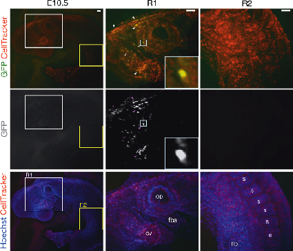 Representative images showing widespread integration of GFP-labelled human naive iPS-derived cells into different locations in the anterior part of an E10.5 mouse embryo. Hoechst and CellTracker were used for counterstaining. The first column shows the whole embryo (z-stack interval 30 μm, 18 focal planes in total). The second column shows a zoom in images focusing on the head region (white square R1) where the human iPS-derived cells (GFP-positive cells) are pointed out (arrowheads, z-stack interval 20 μm, 11 focal planes in total). The third column shows the posterior part of the embryo (yellow square R2) where no GFP-positive cells were detected (z-stack interval 20 μm, 9 focal planes in total). Squares in the first two images in the second column represent the area shown in the insets at the corner of each image. ov, optic vesicle; op, otic pit; fba, first branchial arch; flb, forelimb bud; s, somite. Scale bars, 50 µm.