
Method Specification

Courtesy of Prof. Milko van der Boom’s group
X-ray tomography is ideally suitable for non-destructive three-dimensional visualization and analysis of a wide range of samples, from biological tissues to synthetic materials. Tomographic datasets are obtained by imaging a rotating sample positioned between an X-ray source and an X-ray detector. A set of at least several hundred projections is used for volume reconstruction, which allows the visualization of the inner details. Micro-CT is nondestructive and therefore has no impact on the ability to use the sample for further analysis. Sample dimensions can range from tens of micrometers to several centimeters, with spatial resolution as good as one micrometer.
Observation of low-absorbing materials is possible, sometimes by staining the sample (for example, biological tissues) with solutions containing heavy elements, and sometimes without staining, by using techniques such as propagation phase contrast imaging.
Our facilities allow micro CT imaging at room temperature and also at low temperatures (down to 7 oC) and under mechanical loadings in forms of tension, compression, and three-point bending.
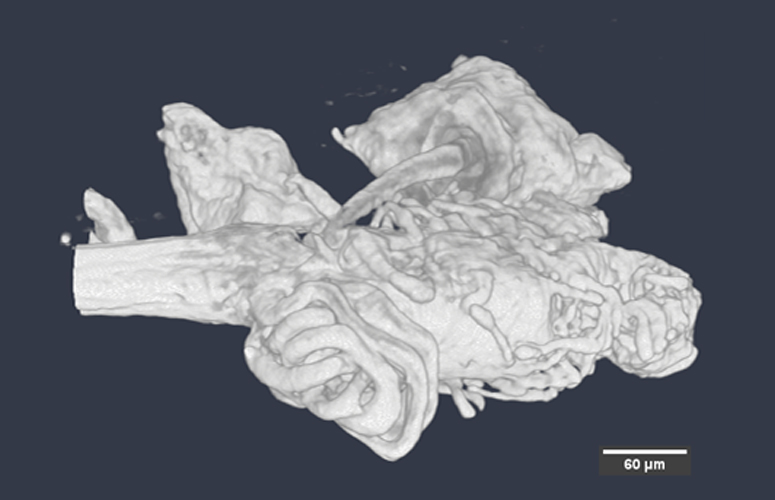
Part of the sexual apparatus of Drosophila Melanogaster
Staff Contacts
-

Dr. Xiaomeng Sui
Staff Scientist -

Dr. Vlad Brumfeld
Consultant