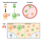
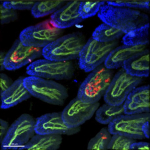
Research

macrophages (red) in small intestinal villi, vasculature (green)

Macrophages - brain / lung / intestine / fat tissue

Tissue macrophages are strategically positioned throughout the organism. As professional phagocytes, they ingest and degrade debris and foreign material, including pathogens, and orchestrate inflammatory processes. Macrophages can be generated from three distinct sources: two early, currently considered transient, hematogenic waves initiated in the yolk sac that seed tissues prenatally, and a pathway involving hematopoietic stem cells and mature blood monocytes, that persists throughout adult life. Most macrophage compartments are established prenatally, and develop independently from each other in their respective host tissue. These cells locally acquire distinct epigenomic and transcriptomic identities, through the imprint influence of the respective microenvironment. While recent studies have revealed critical contributions of tissue macrophages to organ development and homeostasis, specific involvement of discrete subpopulations in the maintenance of the healthy balanced steady-state remain however to be elucidated. Using the mouse as a model, we aim to define functional contributions of specific tissue macrophages to physiology and pathophysiology.

Monocyte ontogeny dictates tissue macrophage identity

Monocytes

Monocytes are mononuclear phagocyte precursors that circulate in the blood and comprise two major subsets. Classical monocytes (CM) are highly dynamic and short-lived. Under homeostasis, these cells give rise to selected tissue resident macrophage populations in barrier tissues, as well non-classical monocytes (NCM), that patrol the inner vessel wall and surveille it for integrity. Following injury and pathogen challenges, CM are recruited to the site of inflammation and complement functions of tissue resident mononuclear phagocytes. Interestingly, emerging evidence suggests that CM themselves are heterogeneous. Differential functions and fates of these  GMP- and MDP- derived monocytes, as well as distinct contributions to tissue homeostasis and pathologies remain however incompletely understood. 

something completely different ..... Studying fungal commensals

We are 'holobionts' like lichen or corals - multispecies superorganisms that consist of animal and symbiotic microbes, on which normal physiological functions depend. Mycobiota are a critical part of the gut microbiome, but host-fungal interactions and specific functional contributions of commensal fungi to host fitness remain incompletely understood. We recently found by serendipity that the fungal commensal Kazachstania heterogenica var. weizmannii could prevent Candida albicans colonization of murine intestines, and even significantly reduce the commensal C. albicans burden in pre-colonized animals, K. weizmannii thereby mitigated candidiasis development. Emerging data from other laboratories have confirmed the dominance of Kazachstania strains over other intestinal mycobiota.

We now aim to define the mechanism underlying the robust fungal competition and explore if competitive fungal commensalism, or associated circuits, can be harnessed for the management of C. albicans-mediated diseases. 

Dendritic cells

Classical Dendritic cells (cDC) are migratory myeloid cells with an unrivaled potential to stimulate naïve T cells. Adoptive transfer experiments have established that cDC are derived from dedicated DC precursors which differentiate in tissue context into distinct DC subsets, including XCR1+ (CD8a+) cDC and CD11b+ DC. Functional differences between these subsets remain incompletely understood, as is their relation to monocyte-derived DC that complement the cDC compartment during inflammation.