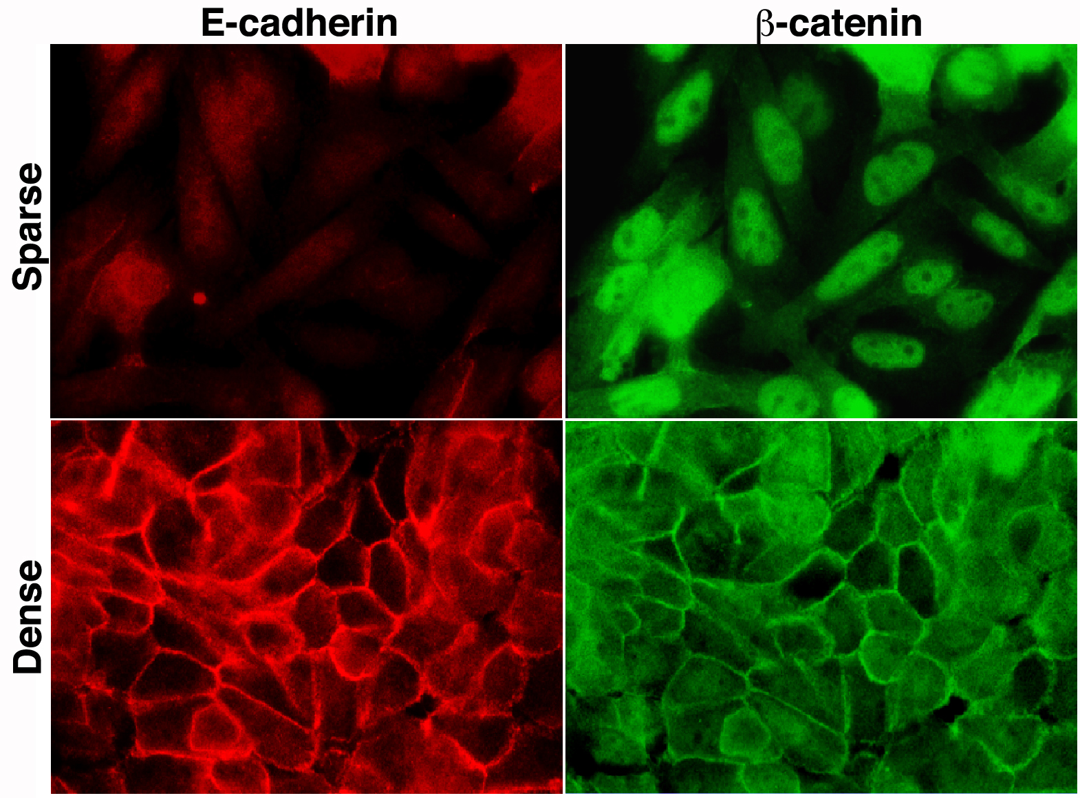
Epithelial-mesenchymal transition (EMT) is a key cell biological process displayed in multiple tissues of the developing embryo. Co-opting the EMT process by cancer cells, and the associated changes in cell adhesion properties are considered major routes for tumor progression. More recent in vivo studies in cancer tissues and circulating tumor cell clusters suggest a stepwise EMT process rather than an “all-or-none” transition during tumor progression. We are studying the mechanisms underlying the plasticity in adhesion properties and in the EMT process displayed by colon cancer cells (see the figure below) hoping to understand its role in colon cancer metastasis.

Human colon cancer cells display a reversible epithelial to mesenchymal (EMT)-like behavior by changes in culture conditions. When grown in sparse cell culture the cells are elongated (more fibroblastic), display strong nuclear β-catenin localization and a very low level of diffuse cytoplasmic E-cadherin. At high cell density, the cells organize into cuboidal epithelial colonies with extensive E-cadherin expressed at cell-cell contacts and with β-catenin co-localizing mostly in these cell-cell adherens junctions together with E-cadherin. Nuclear β-catenin staining is greatly reduced under these conditions. Two days culture under these different conditions can shift the phenotype from a more epithelial to a more mesenchymal (back and forth), demonstrating the plasticity in the epithelial phenotype.


