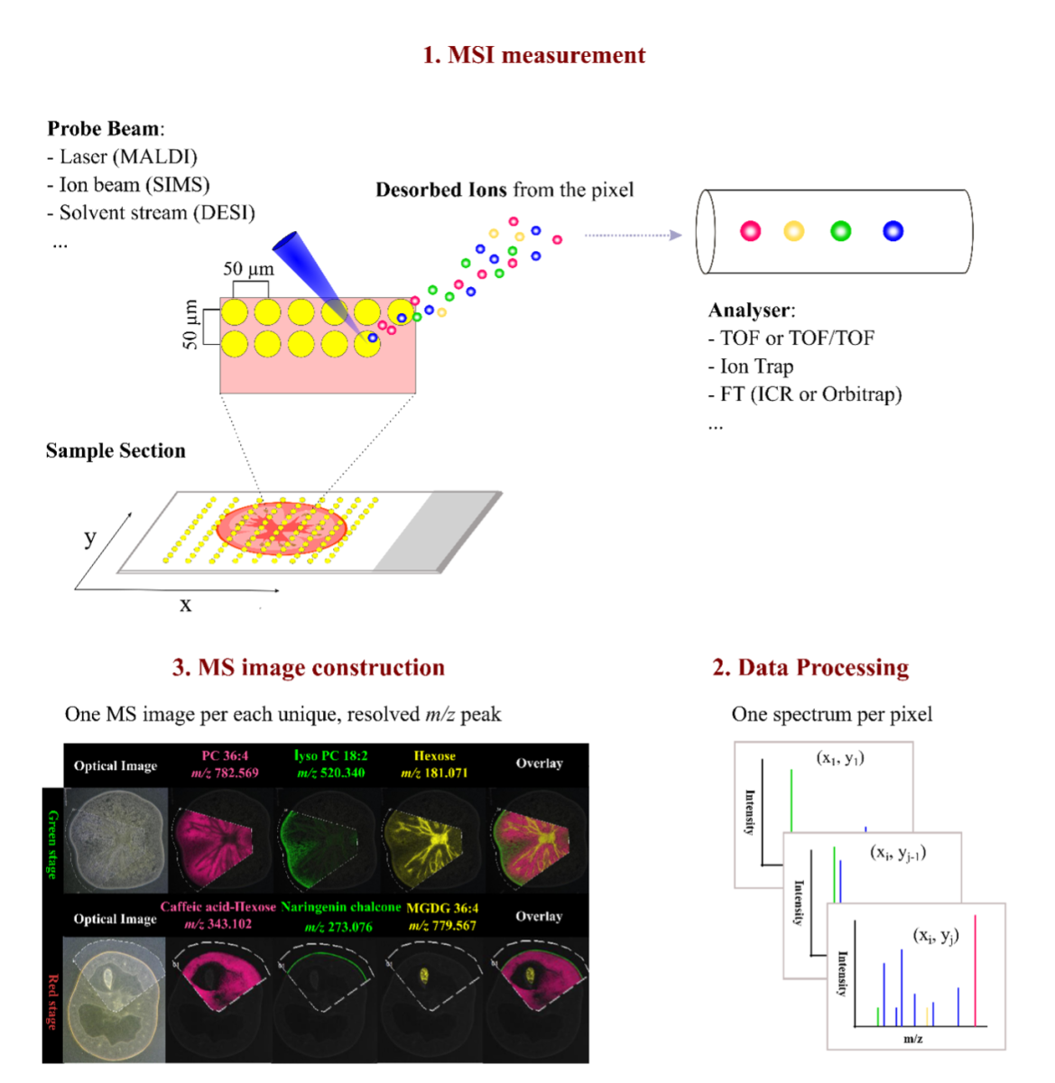
Mass spectrometry imaging is a technique that allows visualization of the distribution of small molecules on a biological sample or section.
In general, molecules are ionized in a spatially resolved manner and detected using a mass analyzer.
The unit offers service for Matrix Assisted Laser Desorption Ionization (MALDI) imaging, Secondary Ion Mass Spectrometry (SIMS) and sample preparation for both techniques.
The two technologies are complementary analysis methods in order to obtain a broad picture of the spatial organization of the metabolome of a biological sample. Various molecule classes are detectable, including primary and secondary metabolites, lipids and peptides.
MALDI imaging
MALDI imaging is a 2D technology that uses a UV laser for ionization of target molecules. Therefore, thin sections of a tissue are coated with chemicals (Matrix) that are ionized by the laser irradiation. During this process, the charge is transferred onto the target molecules, which are detected in the mass spectrometer. The lateral resolution is dependent on the laser focus. Molecule distributions can be resolved to a pixel size of 10 µm. Dependent on the mass spectrometer coupled to the MALDI source mass resolution of up to 1.000.000 can be achieved. (for more details see Services)
SIMS
Secondary ion mass spectrometry uses focused ion beams to bombard the surface of samples. Thereby secondary ions (the analytes) are formed and can be detected via mass spectrometry. The technology enables analysis with a lateral resolution down to 50nm pixel width and dependent on the mass analyzer and measurement mode used a mass resolution of 240.000. Since ions are produced without the need of ionization aids (Matrix) samples can be analyzed in 2D and 3D. (for more details see Services)
Sample preparation
Both, MALDI imaging and SIMS are vacuum techniques hence samples are analyzed dehydrated. The unit offers all equipment needed for preparation of “fresh” biological samples for the analysis, including embedding, cryo-sectioning, soft freeze-drying and transfer of samples on suitable sample holders. Furthermore, the unit is equipped with a HTX sprayer for MALDI matrix application, on slide protein digest and on slide derivatization. (for more details see Services)
Data analysis
The unit offers full analysis of acquired data, including visualization of selected mass traces, mass spectra interpretation and statistical analysis.

Figure: General workflow of a typical MSI procedure. (1) A thin-section sample is mounted onto a glass slide, coated with or without matrix. By using a laser (MALDI), ion beam (SIMS), or charged solvent stream (DESI), ions are generated at each spot (pixel) which are then detected by a mass spectrometer. (2) Mass spectrum is produced from each spot (pixel). (3) After the acquisition, ion intensity of each unique, resolved m/z peak can be extracted from each spot and plotted as a scaled false color heat map-like image, termed MS image or ion image. As exemplified here, single MS images for several different metabolites, such as phosphatidylcholine (PC) 36:4 and naringenin chalcone, are generated to represent their spatial intensity distributions in two different tomato fruit sections. In addition, MS images can be overlaid to compare their distributions.
