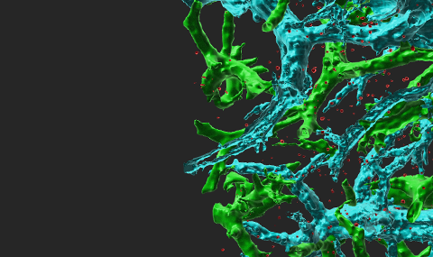
Infection of mCherry encoding influenza virus in the lung airways
Infection of mCherry encoding influenza virus in the lung airways
3D image of lungs infected with the virus imaged by light sheet microscopy (LSM) 4 days post infection. The influenza infected airways are depicted in red. Green: auto fluorescence of the lung stroma. Note the high specificity of the virus towards the airways and the large peribronchial vessel (green) surrounding the virus infected bronchi (red).


